
For several years now, the development of rapid, sensitive, portable and inexpensive diagnostic techniques has been attracting growing interest in the healthcare sector, in both primary care and emergency medicine. The researchers in question have previously demonstrated the proof-of-concept of a patented microfluidic biochip based on Giant Magneto-Resistance or GMR sensors (developed at the SPEC/LNO) arranged on both side of a microfluidic channel that enables individual dynamic detection of biological targets magnetically marked by beads functionalized (at the CEA-Joliot/LERI) by antibodies directed against the target of interest
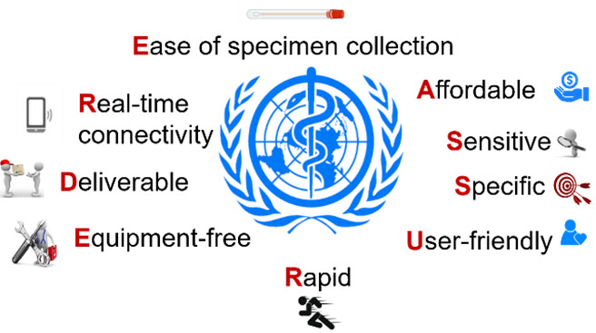
This study was continued by evaluating a number of criteria that define a field diagnostic test (REASSURED criteria defined by the WHO, figure 1) on a murine myeloma cell line in semi-complex matrix (culture medium).
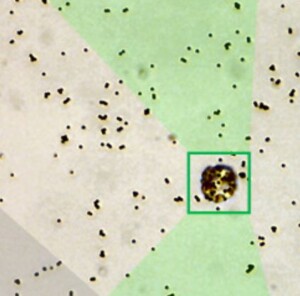
Although of no clinical interest, this biological target has the advantage of being non-pathogenic (and can therefore be studied outside L2 or L3 high-risk laboratories), easily cultivated in vitro , and of being large enough in size (diameter between 7 and 10 µm) to allow visual verification of its magnetic labelling using a simple optical microscope (figure 2B). The results obtained, presented in figure 2A, are very encouraging and competitive with existing techniques
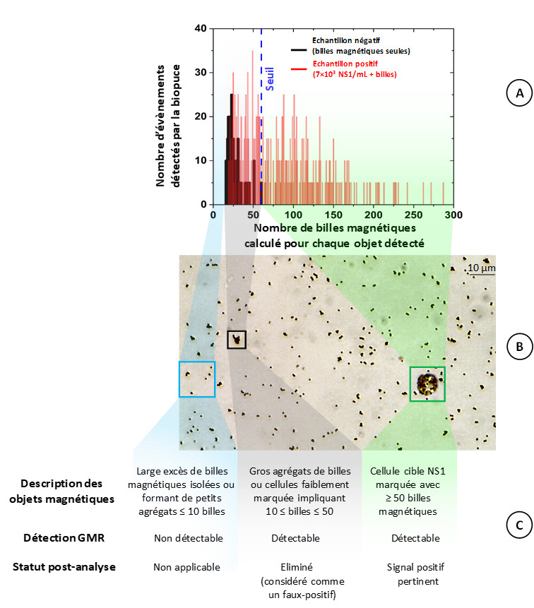
A – Graphical representation of magnetic objects detected by the biochip.
B – Optical microscope photograph of the positive sample shown in A.
C – Description of the magnetic objects present in photo B and their detection and processing by the biochip.
Finally, under a “POC in Labs” contract from the Université Paris-Saclay in collaboration with CEA-Iramis/LETS, the device is being miniaturized for easy transport (figure 3). A new, much lighter Halbach-type magnet with an internal diameter of 10 cm (into which the biochip for polarizing the magnetic beads is inserted) has been designed at SPEC/LNO, and cases for the electronics and transport of the device are being manufactured at the laboratory.

By adjusting some parameters, such as the size of the microfluidic channel of the biochip and the type of antibodies coupled to the magnetic beads, the technology can be adapted to a wide variety of biological targets, demonstrating its ease of implementation and versatility. In collaboration with the Laboratoire Bactériologie et Hygiène at Hôpital Béclère, the team is now focusing on the detection of bacteria and yeasts of interest in the diagnosis of sepsis (or septic shock). To this end, the researchers have joined the IHU-SEPSIS (Institut Hospitalier Universitaire, of which CEA is a partner) created in 2023, which aims to halve sepsis-related mortality and sequelae within ten years. This disease is caused by an uncontrolled immune response to an infection, which then spreads through the bloodstream, potentially leading to the patient’s death. They are taking part in “workpackage 7” of this IHU, the subject of which ranges from the development of innovative diagnostic methods to the design of a rapid multiplex test demonstrator.
References
[1] Proof of concept of a two-stage GMR sensor based lab-on-a-chip for early diagnostic tests, Lab Chip, 2022, 22, 2753-2765.[2] Innovative and sensitive detection of a cancer cell line using a GMR sensor-based biochip prototype for diagnostic purposes, Sens. Diagn. 2025,4, 596-608.
See previous highlight:
CEA contact:
- Guénaëlle Jasmin (SPEC/LNO)
- Agathe Trillat (SPEC/LNO)
- Cécile Féraudet-Tarisse (Joliot/SPI/LERI)