
Air Force Research Lab. AFRL 01 July 2020 – 30 June 2023 Grant number FA8655-20-1-7039
Air Force Office of Scientific Research AFOSR
Competition IOE – European Office of Aerospace Research and Development (EOARD)

Advances in materials science offer a wide range of options for exploring new physical properties, particularly under extreme conditions. Among these, the strong-field regime is a leading area of research. Beyond a critical electric field, any solid emits electrons via the tunneling effect, a quantum-mechanical phenomenon. In terms of applications, this production of electrons via field emission may be desirable or, conversely, undesirable.
Photocathode-type electron source technology falls into the first category. The goal is to generate high current densities and high currents. Target applications include, for example, particle accelerators and radiofrequency devices. In stark contrast, this same phenomenon can be detrimental to the proper functioning of many components. The occurrence of breakdowns in high-gradient accelerators falls into this category. Here, the electrons produced are responsible for localized degradation that causes irreversible structural damage to the device. Thus, understanding electron emission phenomena under high-field conditions is an important topic.
The goal of the POEEMS project is to address fundamental questions related to electron emission from a surface subjected to an extreme electric field. We will study the emission physics of electron-emitting materials under real operating conditions using a multidimensional (space, energy, time) approach based on photoelectron emission microscopy. PEEM microscopy enables the spatial mapping of the distribution of photoemitted electrons at the mesoscopic scale. It allows us to highlight local variations in emission efficiency in relation to the material’s surface properties. PEEM microscopy is a technique sensitive to the kinetic energy of charge carriers. It will establish the energy distribution curves of electrons at the mesoscopic scale, providing a signature of the physical mechanism at play. PEEM microscopy relies on a pulsed laser source, which enables the tracking of electron emission dynamics on a short time scale of the order of hundreds of femtoseconds (1 fs = 1.10⁻¹⁵ s).
All of these measurements will contribute to our understanding of the electron emission characteristics of these new materials and provide feedback for their growth and modeling.

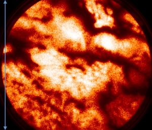
Field-emission PEEM image of a carbon nanotube fiber. The signal originates from the surface of the nanotubes. Field of view: 6 ± 1 µm (blue arrow); applied static electric field: 0.00448 V/nm; logarithmic histogram