
Magnetic Resonance Imaging (MRI) is usually performed with nuclear spins polarized under a strong field of several Tesla. MRI can also be obtained at fields in the millitesla range, provided a detector of sufficient sensitivity is available. The LNO SPEC team is developing such detectors, consisting of a superconducting loop and a giant resistance magnetic sensor.
The precession frequency of the total magnetic moment depends on the strength of the polarizing field (for the hydrogen atom – proton: 12 MHz at 3 T; 43 KHz at 1 milliTesla). As a source of contrast, observable relaxation times for the components parallel and perpendicular to the polarizing field are very different at high and low fields. Low-field MRI is therefore a complementary technique to high-field MRI.
The development of ultra-sensitive magnetic field detectors has enabled the IRAMIS/SPEC team to develop a low-field MRI (< 10 mT), which, in addition to its low cost and ease of use, can be used to complement original diagnostics (prostate cancer, stroke, monitoring of very premature births, correlations with magneto-encephalography, etc.).
Magnetic Resonance Imaging (MRI) is widely used for clinical diagnosis. It is based on the detection of radiation emitted during proton spin relaxation (return to equilibrium) after resonant radiofrequency excitation. It provides a contrasted image of different tissues, based on their influence on relaxation. Not very sensitive, the technique uses strong magnetic fields, of the order of 1.5 T or 3 T, which enable a sufficient signal-to-noise ratio to be achieved for good image quality. However, to date, the technique has not been able to detect quantitative differences between healthy tissue and certain tumors, as in the case of prostate or lung cancer. For this reason, several research teams have started work on ultra-low-field MRI, which, although less resolved, could provide the necessary complementary information, with a much more accessible and inexpensive measuring device.
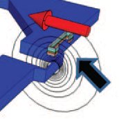
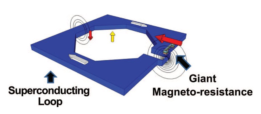
Working at very low fields requires magnetic sensors that are highly sensitive at low frequencies. In the laboratory, a new type of magnetic sensor, called a mixed sensor, has been developed. It is composed of a superconducting loop acting as a flux-field transformer and a giant magnetoresistance (GMR) sensor (figure 1). They enable a sensitivity level of around 100 fT/√Hz (10-15 T/√Hz), over an area of around 1 cm2. In addition, they are extremely robust to the RF pulses used, as well as to external static fields.
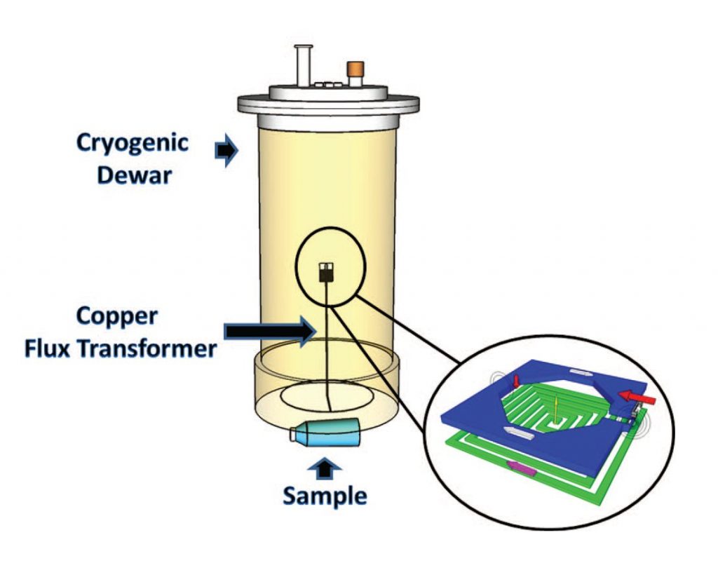
1 cm2 is not, however, a large enough surface area for MRI studies of human tissue. One solution to this problem, already used with SQUIDS, is to couple the mixed sensor to a flux transformer in order to increase the fill factor (figure 2).
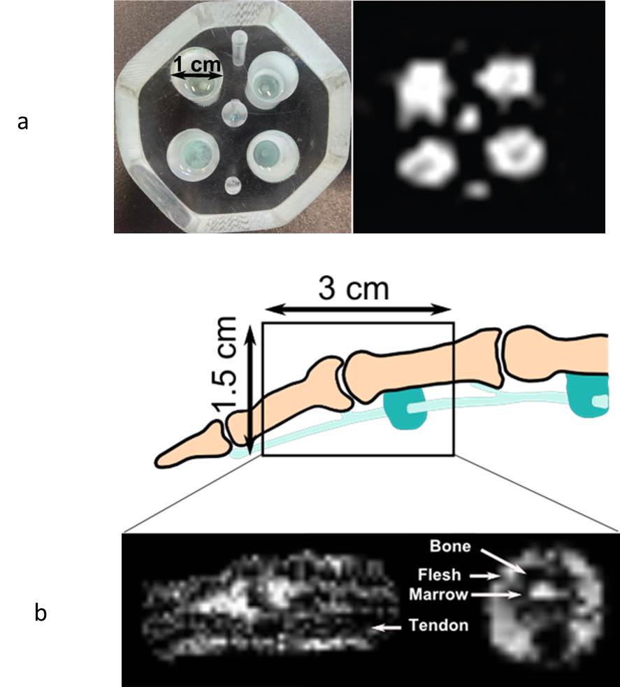
During his thesis at the LNO (SPEC), Quentin Herreros carried out a complete study of this system in order to optimize its gain. In addition, to test its robustness in real-life conditions, the system was incorporated into a compact MRI device, comprising a cryostat and a spectrometer capable of working on a volume of 5 x 5 x 5 cm3 with fields ranging from 1 to 8 mT. At the same time, several MRI imaging acquisition sequences were integrated and adapted to very low field conditions. The system’s excellent reliability has been demonstrated by molecular dynamics studies (using relaxometry) on common liquids and 2D MRI image acquisition on common liquids (figure 3).
Des images IRM in vivo d’un doigt ont également été obtenues à faible champ (7 mT) mais en utilisant une bobine accordée. Pour obtenir les mêmes images avec le capteur mixte, il est nécessaire de modifier le dispositif cryogénique afin de réduire la distance entre le transformateur de flux et l’échantillon mesuré.
En se basant sur ces résultats, un système permettant de réaliser une image d’une tête entière ou d’une partie du corps a été réalisé et est en cours de tests.
References:
[1] Very low field magnetic resonance with spintronic sensor,
Q. Herreros, H. Dyvorne, P. Campiglio, G. Jasmin-Lebras, A. Demonti, M. Pannetier-Lecoeur et C. Fermon, Rev. Sci. Instrum. 84, 095116 (2013)
[2] “IRM à très bas champ magnétique / Very low field MRI”, PhD thesis, Quentin Herreros Université Paris-Sud (2013).
[3] Low field MRI with magnetoresistive mixed sensors
N Sergeeva-Chollet, H Dyvorne, J Dabek, Q Herreros, H Polovy, G Le Goff, G Cannies, M Pannetier-Lecoeur and C Fermon, 2011 J. Phys.: Conf. Ser. 303 012055.
Contacts IRAMIS/SPEC: Claude Fermon, Guénaëlle Jasmin-Lebras et Myriam Pannetier-Lecoeur.