

Researchers from the LNO at SPEC (CEA-CNRS joint research unit), in collaboration with teams from the Ernst Strüngmann Institute (ESI), demonstrate for the first time the possibility of recording in vivo the magnetic signature of single-neuron action potentials, opening a new avenue for studying neural circuits.
Measuring neuronal activity has so far relied mainly on electrical signals. Yet, these currents inherently generate magnetic fields, which in principle carry complementary information and are less affected by the surrounding biological tissue. While such magnetic signals can be measured at the macroscale using magnetoencephalography, their detection at the level of a single neuron in vivo has remained a major challenge, due to their extremely low amplitude and the associated technological constraints.
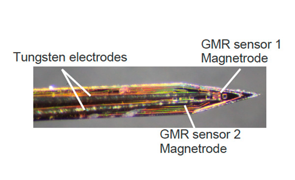
To address this challenge, the researchers developed miniaturized sensors based on giant magnetoresistance (GMR), integrated into microprobes that can be positioned in close proximity to neurons. These sensors provide sufficient sensitivity in the nanotesla range. Estimates indicate that an action potential generates a magnetic signal of approximately 1 nT at a distance of 1 µm and about 50 pT at 10 µm, within a typical bandwidth of 500 to 5,000 Hz, highlighting the extreme difficulty of such measurements.
The key contribution of this study is the experimental demonstration, after applying spike-sorting methods to isolate single units, of magnetic signatures correlated with neuronal action potentials. Across the recordings, a small number of units showed significant correlations between electrical and magnetic signals, with measured amplitudes on the order of a few nanoteslas. These results constitute a proof of principle for the feasibility of in vivo magnetic recordings of action potentials, although detection remains limited and requires averaging across many events.
Beyond this experimental achievement, the work opens important perspectives for neuroscience. Magnetic signals, which are largely unaffected by tissue properties and inherently provide directional information, could complement electrical recordings to improve the characterization of neural circuits. The study also highlights the need for further improvements in sensor sensitivity and probe design to enable more systematic measurements and, ultimately, the direct detection of individual action potentials without averaging ; paving the way for new approaches to probing brain activity.
Référence
“In vivo magnetic recording of single-neuron action potentials”, Frederike Klein, Patrick Jendritza, Chloé Chopin, Mohsen Parto-Dezfouli, Aurélie Solignac, Claude Fermon, Myriam Pannetier-Lecoeur, Pascal Fries, Journal of Neurophysiology 134 (2026).
Collaboration
Contact CEA
Myriam Pannetier-Lecoeur, Laboratoire Nano-Magnétisme et Oxydes – LNO, Service de Physique de l’État Condensé – SPEC, CEA-IRAMIS.