Researchers from the Laboratoire Léon Brillouin (LLB), in collaboration with Synchrotron SOLEIL, the Laboratory of Microbial Ecology of the Rhizosphere – LEMiRE du BIAM, and Sanofi have shown that certain RNA molecules can insert into the membrane of outer membrane vesicles (OMVs) released by Gram-negative bacteria. Using an original spectroscopic approach, they reveal a previously unrecognized localization of RNA associated with these vesicles, providing new insights into the potential mechanisms underlying communication between bacteria and host cells.
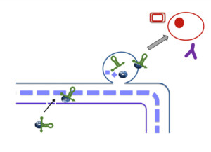
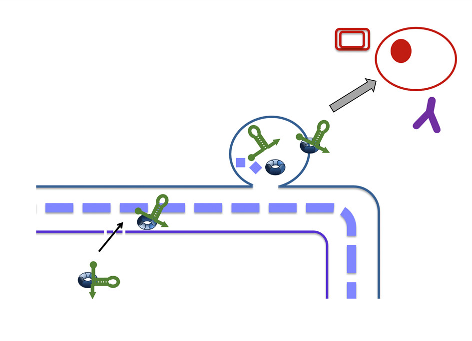
Gram-negative bacteria naturally release outer membrane vesicles (OMVs), nanoscale membrane-bound particles that transport a wide range of biomolecules involved in interactions with their environment. Although OMVs are known to carry proteins, peptidoglycan and RNA molecules, the precise localization of these RNAs has remained unclear. Until now, RNAs were generally thought to reside mainly within the OMV lumen. Whether RNAs could be found and inserted into the OMV membrane itself, however, remained an open question, with important implications for understanding how these molecules may interact with other cells.
To address this question, the researchers implemented an innovative application of Synchrotron Radiation Oriented Circular Dichroism (SR-O-CD), a spectroscopic technique capable of probing the orientation of molecules associated with biological membranes. Applied for the first time to bacterial outer membrane vesicles, this approach makes possible to distinguish a simple RNA-membrane interaction from genuine insertion into the lipid bilayer. The experiments show that RNA insertion depends on the C-terminal region (CTR) of the Hfq protein, whose ability to interact with cytoplasmic membrane had previously been demonstrated by the authors. In the presence of this domain, the polyadenylated RNA model used in this study (rpsO-polyA) inserts into the OMV membrane, whereas no insertion is detected in its absence.
These findings support a new model for the organization of OMV-associated RNAs. They suggest that RNAs associated with OMVs may display a dual localization, being present both within the vesicle lumen and, for some species, within the vesicle membrane. The study also shows that not all RNAs behave similarly: membrane insertion depends on the sequence of the RNA examined and on its interaction with Hfq, pointing to a selective rather than passive mechanism. Such dual localization could influence how these RNAs are presented to host cells or to other bacteria, although this hypothesis remains to be investigated.
Beyond these findings, the study provides new insight into the molecular organization of bacterial outer membrane vesicles and introduces an innovative approach for investigating RNA insertion into complex biological membranes. While the in vivo functions of these membrane-associated RNAs remain to be established, this work opens new avenues for exploring the roles that OMV-associated RNAs may play in communication between bacteria and host cells. It also provides a valuable experimental tool for investigating these still poorly understood mechanisms.
Reference
RNAs associated with bacterial outer membrane vesicles: structural insights into surface composition,
Mosca, K., F. Turbant, W. Achouak, F. Wien, and V. Arluison. 2026. Journal of Extracellular Vesicles15, no. 6: e70306.
Collaboration
- Équipe matière molle et Biologie – MMB du Laboratoire Léon Brillouin, UMR 12 CEA-CNRS
- Laboratoire d’Écologie Microbienne de la Rhizosphère – LEMiRE, BIAM UMR 6191 CEA-CNRS-Aix-Marseille Université
- Ligne DISCO, Synchrotron SOLEIL
- Centre de Recherche & Développement de Sanofi Marcy-l’Étoile
Contact
- Véronique Arluison, Researcher at LLB and Université Paris Cité.