Observing the ferroelectric texture of nanocrystals remains a challenge, due to the size of the objects, and of the modulation of their properties, and the weakness of the signal.
Teams from French laboratories, including SPEC/LEPO, and American laboratories have pooled their know-how to study experimentally and theoretically the ferroelectric structure of barium titanate (BTO) nanocrystals.
Their results, published in the journal ACS Nano, pave the way for new designs in optical or thermal nanosensors and their potential applications in biology.

Ferroelectric materials exhibit unique properties such as high dielectric permittivity and ferroelectric hysteresis, which are the basis for a wide range of technological applications (electronics, optics, etc.). Although the vast majority of studies have focused on thin films of these materials, recent advances have enabled the fabrication of ferroelectric nanocrystals, for which original polarization textures can be obtained and deserve to be studied and better understood.
Following the objective of designing biocompatible optical sensors from barium titanate (BTO) nanocrystals (ANR UFO project), a collaboration between French and American teams studied nanocrystals synthesized in cubic form, with an average size of around 160 nm. The ferroelectric “texture” of these nanometric objects was probed using piezoelectric force microscopy (PFM). This measurement technique uses a tip as a local probe in contact with the sample, and takes advantage of the multiferroic character of the crystals, both ferroelectric and piezoelectric. In parallel, simulations of the equilibrium electrical polarization distribution on a nanocrystal and of the PFM response were carried out using a phase field model (FERRET module).
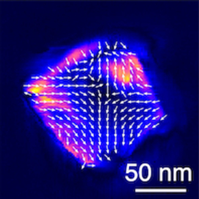
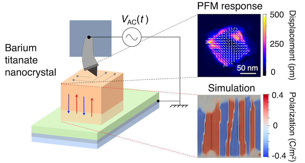
Measurements reveal that the facets of all the nanoBTOs studied have a PFM response, showing local in-plane deformation as a function of the electrical voltage modulation applied to the tip. Phase-field modelling helps explaining this observation: it reveals that the equilibrium polarization in volume, consisting of alternating up- and down-oriented domains along one of the crystallographic axes of the BTO tetragonal phase (blue and red areas in the simulation figure), rotates by 90° at the surface (grey areas). The simulation also gives a qualitative account of the deformation fields measured by PFM.
Published in ACS Nano, this work demonstrates the value of the PFM technique and paves the way for the design of optical nanosensors (temperature or electric field measurement sensors, in particular) based on nanoBTOs doped with rare-earth ions.

Electrical polarization of a ferroelectric nanocrystal: study by piezoelectric force microscopy (PFM) and phase-field model (simulation).
Left: stacking of a ferroelectric BTO nanocube (orange) on a thin layer of conductive polymer (green), deposited on a conductive ITO deposit (grey-blue) on a glass slide. The PFM tip probes the top face of the cube.
Right, top: The amplitude (color code) and direction (arrows) of displacement of the PFM tip are measured in response to an alternating potential difference applied between the tip and the underside of the nanoparticle.
Right, bottom: result of electrical polarization simulation (phase field model, FERRET module). The image shows, at the center of the nanocrystal, a domain structure of ferroelectric polarization, directed along a crystallographic axis and with alternating orientations. It also shows the absence of vertical polarization on the lower and upper faces (grey areas), in agreement with PFM measurements. © F. Treussart, C. Paillard and C. Fiorini.
For the optical applications initially proposed, the results of the study also raise the question of the origin of the nonlinear second-harmonic generation (SHG) optical response of individual BTO nanocrystals widely described in the literature. Indeed, given the ferroelectric texture revealed in the simulations, the nanocrystals appear to be made up of core and surface domains oriented at 180° to one another, with an overall polarization compensation. A priori, this should result in the absence of SHG by destructive interference, which is not observed experimentally. Further analyses coupling PFM and SHG will be carried out in the near future to gain a deeper understanding of the structure of these nanocrystals.
Reference:
Ferroelectric texture of individual barium titanate nanocrystals.
Athulya K. Muraleedharan, Kevin Co, Maxime Vallet, Abdelali Zaki, Fabienne Karolak, Christine Bogicevic, Karen Perronet, Brahim Dkhil, Charles Paillard, Céline Fiorini-Debuisschert, and François Treussart, ACS Nano 18 (28) (2024) 18355
Open access: HAL et Arxiv (follow the link)
Post-doct at CEA-SPEC and PhD thesis of Athulya K. Muraleedharan (decembre 2023) : “Study of the ferroelectric texture of individual barium titanate nanocrystals by piezoresponse force microscopy. Application of erbium-doped nanocrystals to optical detection of electrical potential”.
Study carried out as part of the ANR SINAPSE and UFO projects: “Ferroelectric nanocrystals with photon upconversion for optical detection of electrical potential in biological systems”.
Contact CEA-IRAMIS: Céline FIorini, SPEC/LEPO
Collaboration:
- Université Paris-Saclay, Laboratoire lumière-matière aux interfaces (LUMIN), UMR CNRS/ENS Paris-Saclay, Université Paris-Saclay,
- Université Paris-Saclay, Laboratoire Structures, propriétés et modélisation des solides (SPMS), CentraleSupélec,
- Université Paris-Saclay, Laboratoire d’Électronique et nanoPhotonique Organique, Service de Physique de l’État Condensé – SPEC, UMR CEA-CNRS,
- Département de physique de l’Université d’Arkansas aux États-Unis.

