Currents circulating in excitable cells like neurons or nerve fibers may be measured by the radiated magnetic field. At the organ level, these magnetic fields can be detected by non-invasive experiments using highly sensitive magnetometers such as SQUIDS, atomic magnetometers or mixed sensors, the latter using spin electronics. This technique, called Magneto-Encephalography, allows measuring neuronal activity at a millisecond resolution and for collective response of population of typically 10 000 neurons and more. To understand the genesis of the signals obtained in brain areas, it is relevant to investigate the fields generated at the level of one or few cells. This requires small and sensitive field sensors, operating at physiological temperatures, which has long been out of reach from existing technologies.
Spin electronics allow now developing small sized and very sensitive magnetometers, reaching the sub-nanotesla field range on micron-size sensors. These devices operate from low temperature to hundreds of °C, so they can be used at physiological temperature. Furthermore, spin electronics sensors, based on thin film technology, can be deposited on silicon or glass substrates which can be shaped in needle-type devices to allow penetration in tissues with reduced damages.
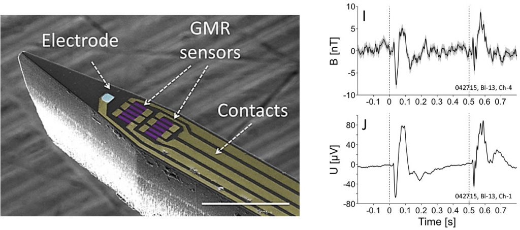
In the frame of the European FET-project “Magnetrodes”, the Nanomagnetism and Oxydes laboratory (LNO) at CEA-Saclay has designed and fabricated magnetic sensors called magnetrodes, as a magnetic equivalent of electrodes, to probe locally the information transmission of excitable cells. These sharp probes contain GMR elements in embodiment compatible with recordings within tissues.
In collaboration with Pascal Fries’team at the Ernst Strüngmann Institute in Frankfurt, Germany, the LNO laboratory has realised the first in vivo experimental measure of the magnetic signature of local field potentials in the cat’s visual cortex. It has paved a new way to a local description of electrical activity, without direct contact to the cell and which allows accessing not only the amplitude of the activity but also its direction of propagation, at any depth within the tissues.

Perspectives of these local magnetic recordings are to target the measurement of single events and of single cell responses, in particular through Action Potential detection which would lead to a spatially and time-resolved mapping of the neural currents running inside neurons.
Reference
Laure Caruso, Thomas Wunderle, Christopher Murphy Lewis, Joao Valadeiro, Vincent Trauchessec, Josué Trejo Rosillo, José Pedro Amaral, Jianguang Ni, Patrick Jendritza, Claude Fermon, Susana Cardoso, Paulo Peixeiro Freitas, Pascal Fries, Myriam Pannetier-Lecoeur, In Vivo Magnetic Recording of Neuronal Activity, Neuron, Volume 95 , Issue 6 , 1283 – 1291.e4 (2017)
Collaboration
- SPEC, CEA, CNRS, Université Paris-Saclay, France
- Ernst Strüngmann Institute (ESI) for Neuroscience in Cooperation with Max Planck Society, Germany
- Instituto de Engenharia de Sistemas de Computadores-Microsystems and Nanotechnology (INESC-MN), Portugal
- Instituto Superior Técnico IST, Physics Department, Universidade de Lisboa, Portugal
- Donders Institute for Brain, Cognition and Behaviour, the Netherlands
Contact CEA
Myriam Pannetier-Lecoeur, Laboratoire Nanomagnétisme et Oxydes (LNO), DRF/IRAMIS/SPEC – UMR 3680 CEA-CNRS


