



For medicines based on RNA or DNA, the chemical composition of these molecules is not the only factor that matters: their shape can also affect their stability and how they function. Yet DNA and RNA can fold into different structures, several of which may coexist in the same sample, making them difficult to analyse. Researchers have developed NA-CDQuant, a UV light-based method that estimates the proportion of each structure present. By making these structures easier to characterise, the tool could contribute to the development and quality control of RNA- and DNA-based treatments.
DNA is often pictured as a double-helix. Yet this familiar structure is not the only form it can take. Like RNA, DNA can fold in different ways, forming loops or more complex arrangements. These structures are not fixed: they can change with temperature, their chemical environment or interactions with other molecules. They play an important role in biological processes, including the regulation of gene expression. To better understand these processes, scientists need to identify the different structures that DNA and RNA adopt and determine their relative proportions. This becomes challenging when several structures coexist and their signals overlap during measurements.
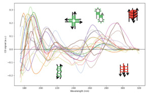
To address this challenge, researchers from the Laboratoire Léon Brillouin – LLB (Joint unit CEA–CNRS) collaborated with Synchrotron SOLEIL, Sanofi, Université Paris Cité, Aarhus University (Denmark) and the South Dakota School of Mines and Technology (United States). They used a technique known as circular dichroism spectroscopy, which measures the difference in how molecules absorb left- and right-circularly polarised ultraviolet light. This difference depends, in part, on how the molecules are arranged in space. It produces a characteristic signal, known as a spectrum, which provides information about their structure. However, when several structures are present in the same sample, their signals overlap and become difficult to interpret. To distinguish them, the researchers analysed 225 DNA and RNA spectra, including measurements collected at Synchrotron SOLEIL in France and at the ASTRID2 synchrotron at Aarhus University in Denmark. From these data, they identified 18 reference signatures representing different families of structures and developed NA-CDQuant, an algorithm capable of estimating the contribution of each structure to the measured spectrum.
To assess the method’s performance, the team tested it on nine DNA structures and two RNA samples whose structures had previously been characterised using other techniques. The results show that NA-CDQuant can estimate the proportions of different structures, although its accuracy depends on their complexity. When several parts of the same molecule interact, accounting for these interactions improves the estimates. By contrast, for mixtures of two distinct DNA structures prepared in known proportions, a simpler analysis is sufficient to recover their relative contributions. The method therefore goes beyond simply identifying a structure: it provides quantitative information about the structural composition of a sample.
These findings nevertheless require some caution. NA-CDQuant does not produce a direct image of the molecules. Instead, it estimates the presence of different structures from their spectral signatures. Some of these signatures can be similar, occasionally leading to incorrect assignments, particularly in more complex molecules. The researchers also show that measurements using synchrotron radiation, which provides access to a broader range of wavelengths, improve the accuracy of the results. The method can still be used with conventional laboratory instruments, although with lower accuracy.
By making it easier to study the different structures adopted by DNA and RNA in solution, NA-CDQuant will help scientists better understand how these molecules fold and respond to changes in their environment. Future work will focus on expanding the reference library and extending the analysis to more complex or chemically modified structures. In the longer term, the method could also have medical relevance. In RNA- and DNA-based treatments, molecular structure can influence stability and other properties. A method capable of detecting structural changes more effectively could therefore help assess the quality and consistency of manufactured products, or investigate the conditions that favour their stability. Such applications still need to be developed and validated, but they illustrate how a better understanding of the molecules involved in living systems support pharmaceutical research.
Reference
K. Mosca, S. V. Hoffmann, N. C. Jones, R. R. Sinden, F. Wien, S. Marco et V. Arluison. « NA-CDQuant: Quantitative spectral analysis of nucleic acid structure by circular dichroism spectroscopy ». Proceedings of the National Academy of Sciences (PNAS), 123(41), (2026).
Collaboration
- Synchrotron SOLEIL (France).
- Sanofi (France).
- Université Paris Cité (France).
- Aarhus University (Denmark).
- South Dakota School of Mines and Technology (United States).
Contact
- Véronique Arluison, researcher at LLB.