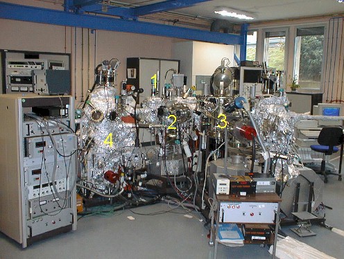
The molecular beam epitaxy technique (MBE) was developed initially for the crystalline growth of the semiconductors. It is an ultra-high vacuum (P Molecular Beam Epitaxy (MBE)
Introduction chamber (1) equipped with a turbomolecular pump.
Preparation chamber(2)
– Gun allowing the ionic bombardment of surfaces (Ar+ ions of energy ranging between 1 and 8 keV).
– Oven allowing a heating until 1800°C by combining the Joule effect until 500°C and the electronic bombardment above. The temperature is measured by a thermocouple and/or an infra-red pyrometer.
– Gas introduction .
– Device of low energy electron diffraction (LEED Riber).
Analysis chamber (3)
– Source X Riber (not monochromatic): Al (hν = 1486.6 eV) or Mg (hν = 1253,6 eV).
– Detection by a hemispherical analyzer (Clam II of VG-scientific).
Growth chamber(4)
– Three Knudsen cells cooled by water allowing the evaporation of metals.
– Gun with ions of low energy (200 to 600 eV).
– Manipulator equipped to heat by Joule effect the substrate until 600°C during the deposition. The rotation of the sample holder is motorized.
– Thickness measurement by a balance with Inficon quartz cooled by water.
– Source of oxidation by monoatomic oxygen plasma.
– Gun RHEED (Staib Instrumente) which uses electrons with a primary energy ranging between 0 and 30 keV. The diffraction pattern is observed on a fluorescent screen. The geometry of the growth chamber allows an incidence of the electrons beam about a few degrees. The images are then recorded by a camera CCD cooled by Peltier effect. The recording and the analysis of the diffraction patterns (LEED and RHEED) are done using the KSA-400 software.
– quadripolar gas Analyser Inficon.
– CMA Auger Spectroscopy (PHI: Physical Instruments).
Setup used at the SPCSI