


MRI (based on Nuclear Magnetic Resonance – NMR)* is a well-known imaging and analysis method used in medicine for clinical diagnosis. The technique is also widely used in chemistry, biology and the study of materials.
A new method, recently developed and based on MRI, explores the local surface magnetization of an object. Applicable to the study of electrochemical processes, the method can be used to improve electricity storage devices for mobile equipments.
This original technique, developed on the NIMBE NMR platform at CEA-Saclay, uses a large-aperture NMR spectrometer (Brücker Avance – Super-Wide-Bore NMR instrument NEO, 7T), advanced MRI techniques and ultra-sensitive custom-built RF sensors. This new analysis procedure has been successfully implemented for the operando study of batteries. In particular, it reveals a series of fundamental electrochemical phenomena associated with lithium ion transport in the positive electrode materials of commercial “pouch” type Li-ion cells.
Among the many MRI methods, a time-efficient purely-phase encoded technique has been successfully adapted by K. Romanenko for an operando analysis of cell batteries [1-7]. In these Li-ion systems, the inhomogeneous intercalation of lithium ions in the battery electrodes introduces local magnetic inhomogeneities that can be unveiled by MRI.
To obtain this type of MRI image, a sensor has been specifically designed: it consists of a thin layer of proton-rich polymer surrounded by a coiled copper ribbon acting as a coil for the NMR excitation. After an initial “blank” measurement, the assembly is placed in contact with the object to be studied (see Figure). As proton resonance frequency variations are a function of the local magnetic field, differential MRI mapping reveals magnetic susceptibility inhomogeneities within the battery under study.

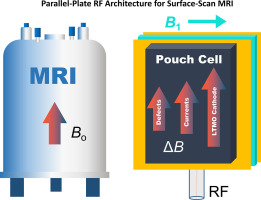
Left: the imaging method uses a 7 T polarizing magnet (B0) with a very wide aperture to insert the measuring device (see text).
Center: a) The surface scanning MRI sensor is composed of thin copper strips (~60 μm thick), surrounding a ~1 mm thick, proton-rich polymer layer. b) Local variations in magnetic susceptibility, depending on Li concentration, within the battery electrode induce an inhomogeneous field (along the B0 direction) within the polymer that is measurable by proton NMR.
Right: during charge or discharge, the Li concentration becomes inhomogeneous within the electrode, then homogenizes by diffusion. Li migrates from the 03-R1 phase to the O3-R2 phase of LixCoO2.
A recent study of the positive LTMO electrodes (L= Li, TM= transition metal, here LiCoO2) of “pouch” cells, used as Li-ion cell phone batteries, illustrates the method. Such imaging enables non-destructive monitoring of kinetic processes within these electrochemical devices with high spatial and temporal resolutions. For these systems, the spatial and temporal fluctuations in magnetic susceptibility that can be detected, are associated with the non-uniform lithiation** of the positive solid electrode, inducing Li+ diffusion and magnetic phase transitions. Another remarkable result of the technique is the ability to track electric current density, via the mapping of lithiation levels on the positive electrode. In addition, it enables real-time monitoring of lithiation homogenization, a slow process that takes place over several hours.

Surface scanning MRI should also enable to explore the magnetic properties of other types of positive electrodes, such as LiNixMnyCozO2 (NMC), LiFePO4 et LiMn2O4, which may exhibit other intercalation reactions. The coupling between the local electrochemical and magnetic properties of these various electrodes opens up a wide range of applications, such as solid-state ion conduction studies that can focus on effects related to intercalant type and concentration, as well as temperature effects.
In the future, these results could also be relevant to the design of more efficient cells, for which the lithiation level can be adjusted locally across the electrode. Another avenue of development could be the on-line control of commercial devices.
* The MRI-NMR method:
MRI is a well-known medical imaging and analysis method based on NMR. The technique is based on the resonant excitation of radiofrequency-induced transitions between nuclear energy levels (Zeeman effect), separated in energy by an intense magnetic field. Transient spatial gradients in the magnetic field induce a frequency shift in the resonances, enabling the information to be localized and the image to be constructed. MRI is widely used in medicine for clinical diagnosis, and for research into chemistry, biology and materials.
**Lithiation: chemical reaction in which lithium atoms are inserted into a material.
References :
[1] Operando magnetic resonance imaging reveals phase transitions driven by nonuniform cathode lithiation in Li-ion pouch cells,
Konstantin Romanenko*, Nikolai Avdievich, Alan Wong, Magali Gauthier, Remith Pongilat, and Alexej Jerschow, Chem. Mater. 35 (22) (2023) 9789.
Other references:
[2] “Distortion-free inside-out imaging for rapid diagnostics of rechargeable Li-ion cells”,
K. Romanenko, A. Jerschow, Proc. Natl. Acad. Sci. U.S.A. 116 (38) (2019) 18783–18789.
[3] “Accurate visualization of operating commercial batteries using specialized magnetic resonance imaging with magnetic field sensing”,
K. Romanenko, P. W. Kuchel, A. Jerschow, Chem. Mater. 32 (2020) 2107–2113.
[4] “Observation of memory effects associated with degradation of rechargeable lithium-ion cells using ultrafast surface-scan magnetic resonance imaging” K. Romanenko, A. Jerschow, J. Mater. Chem. A 9 (2021) 21078–21084.
[5] “Numerical modeling of Surface-Scan MRI experiments for improved diagnostics of commercial battery cells”,
K. Romanenko, A. Jerschow, J. Magn. Reson. Open 10–11 (2022) 100061.
[6] “Surface-Scan MRI diagnostics of Li-ion cells: Boosting the sensitivity with high performance flat RF Sensors”,
K. Romanenko, N. Avdievich, J. Phys. Chem. C 127 (2023) 85−91.
[7] “Unilateral RF sensors based on parallel-plate architecture for improved Surface-Scan MRI analysis of commercial pouch cells”,
K. Romanenko, N. Avdievich, Journal of Magnetic Resonance Open 18 (2024) 100141.
Contact CEA-IRAMIS : Konstantin Romanenko (NIMBE/LSDRM)
Collaboration :
- Université Paris-Saclay, UMR NIMBE CEA-CNRS
- Max-Planck Institute for Biological Cybernetics, Tübingen, 72076, Germany
- Department of Chemistry, New York University, New York, New York 10003, United States.