D. Sakellariou (SCM), G. Le Goff et J.-F. Jacquinot (SPEC)
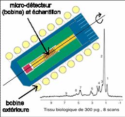
To increase in sensitivity on small samples, it is necessary to approach as much as possible the detector (coils surrounding the sample). So, the difficulty to obtain a spectrum high resolution seems insurmountable since it would be necessary to place the sample in a micro-capillary tube and then spun at several thousand revolutions per second in a stable and reproducible way inside a micro-detector (coil) with an interior diameter of a few hundred microns. To meet this challenge, the CEA team (D. Sakellariou, G. LeGoff and J.-F. Jacquinot) came up with an innovative solution by spinning the micro-detector (coil) and sample in one piece, with power to the detector supplied via induction from an exterior coil, which also enabled the (wireless) transmission of the desired signal. The whole system spins at thousands of revolutions per second, and probably constitutes the world’s fastest set of rotating antennas.

The method proposed is very general and easy to implement in most commercial NMR probes, making it possible to accelerate data acquisition while reducing the minimum quantities of material required. The solution based on rotating micro-coils (MACS or magic angle coil spinning) will be enhanced by the latest miniaturisation techniques and should lead to considerable advances in NMR and micro-imaging (MRMI) as well as metabonomic and biomedical applications (on small biopsies or a few cells) and material science developments. This technique may also pave the way to NMR studies of samples from within containments (e.g. radioactive materials, high-pressure systems).
References:
[1] D. Sakellariou, G. Le Goff et J.-F. Jacquinot,
High-resolution, high-sensitivity NMR of nanolitre anisotropic samples by coil spinning,
Nature 447 (2007) XXX.
[2] D.L. Olson, T.L. Peck, A.G. Webb, R.L. Magin, and J.V. Sweedler
High-Resolution Microcoil 1H-NMR for Mass-Limited, Nanoliter-Volume Samples
Science, 270 , 1967-1970 (1995).
[3] I. Farnan, H. Cho, W. J. Weber,
Quantification of actinide $$-radiation damage in minerals and ceramics,
Nature, 445 , 190-193 (2007).
See also “News and views” de A.S. Edison & J.R. Long
and the “Editor summary” in the same issue of Nature


