
Measurement of the cardiac electrical activity allows following the heart dynamics. Usually measured with electrodes during an electrocardiogram (ECG), this activity can also be studied by monitoring the magnetic component, induced by the circulation of the heart electric currents. This is called “magnetocardiography (MCG). The exquisite sensitivity of the new magnetic sensors made in IRAMIS / SPEC will contribute to the emergence of this technique, which has the advantage of being contactless, therefore requiring no electrodes and allowing a dynamic imaging of the cardiac cycle by a mapping of the signal.

The magnetic signals generated by the cardiac activity are of very low amplitude (tens of picoteslas ~ 10-12 T) and only extremely sensitive magnetometers like SQUIDs [1] (Superconducting Interference Devices) allowed up to now such precise measurements.
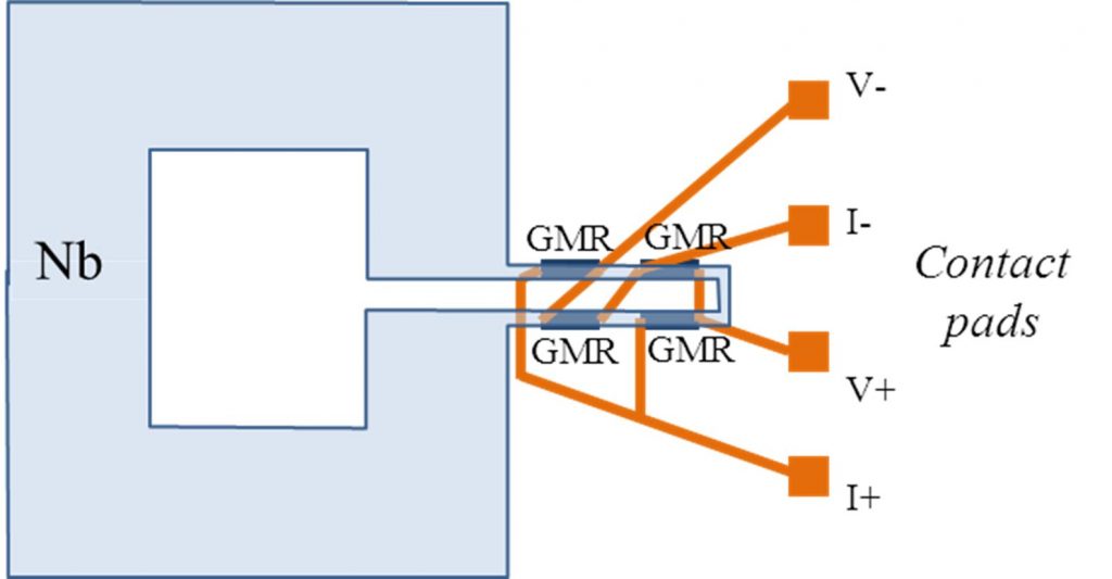
The sensitivity is the key parameter to measure such low signals, and new sensors developed in IRAMIS/SPEC on the principle of spin electronics are used to perform magneto-cardiography measurements. The principle of these sensors is to associate a magnetic field sensor, based on the giant magnetoresistance effect, and a superconducting loop capturing the magnetic flux [1]. With this device, sensitivities of the order of several femtoteslas (~ 10-15 T) are achieved.[1] A SQUID (Superconducting Interference Device) is an electrical circuit made of two branches forming a superconducting ring; each branch includes a thin insulating barrier (Josephson junction). The voltage difference measured across the branches is function of the magnetic flux through the ring, which is quantified.
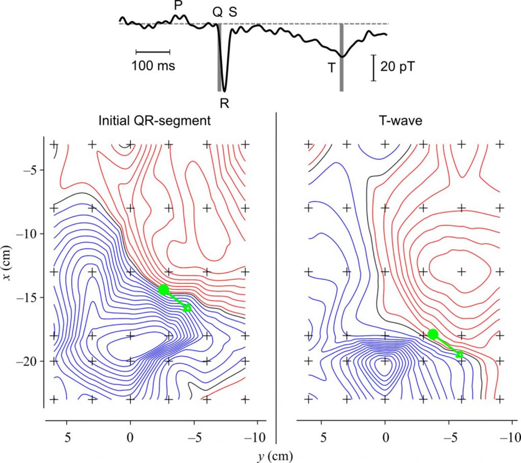
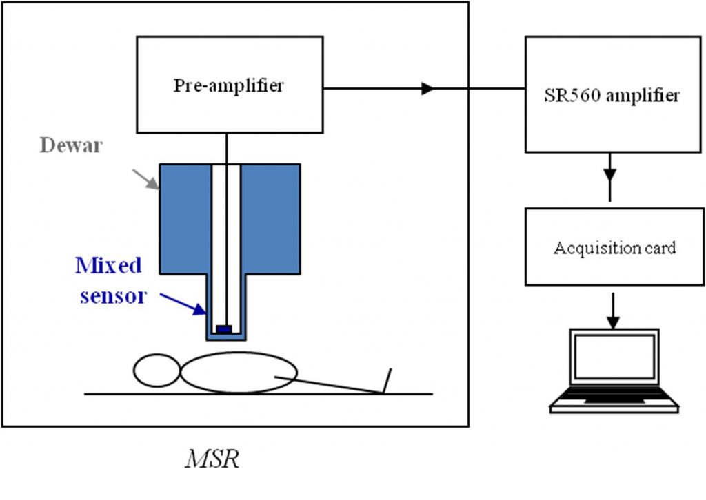
The measurement was performed on healthy adult volunteers in the shielded magnetic chamber in Neurospin (DSV), allowing the elimination of the major part of the external magnetic disturbances. The signal was recorded without contact along a grid positionned above the subject’s torso. From the set of individual measurements, it is possible to plot the isocontours of the magnetic field, and then to locate and get the amplitude of the current dipole associated to the different times of the cardiac cycle.
Following the measurement principle thus established, it is now possible to design a clinical MCG. It could contain a network of 25 to 36 magnetic sensors that will achieve in a very short time, typically one minute, a dynamic image of the electrical activity during the cardiac cycle. In addition, the combination of information received by all sensors would allow reducing the ambient magnetic noise, even in an unshielded environment, by means of the signal analysis techniques developed for magneto-encephalography (MEG).
References:
|
Magnetocardiography with sensors based on giant magnetoresistance, M. Pannetier-Lecoeur, L. Parkkonen, N. Sergeeva-Chollet, H. Polovy, C. Fermonand C. Fowley, Applied Physics Letters 98 (2011) 153705. |
 |
|
[1] Femtotesla magnetic field measurement with magnetoresistive sensors M. Pannetier, C. Fermon, G. Le Goff, J. Simola, and E. Kerr, Science 304 (2004) 1648. |
 |
Contact : Myriam Pannetier-Lecoeur.