N. Barrett et L.-F. Zagonel, Service de Physique et Chimie des Surfaces et Interfaces (SPCSI)
From R. Feynman’s idea at the origin of nanotechnologies “There’s plenty of room at the bottom”, emerged a new discipline: “physics of surfaces”. After the emergence of the near-field microscopies, this topic remains alive today with the current developments of new techniques of electronic imagery.

After the installation of its first Low Energy Electron Microscope (LEEM), the SPCSI continues in this direction and take an active part in the Nanocarac platform associating the “Direction de Sciences de la Matière” and the ” Direction de la Recherche Technologique (DRT)” within CEA to Minatec, around the synchrotron ESRF of Grenoble. In particular, SPCSI offers the skills of its scientists in photoemission spectroscopy.
In July 2006 a new photoemission microscope NanoESCA (Omicron GMBH) was installed. This instrument is able to work in laboratory conditions (bright UV and optimized Al Kα X-ray sources), as well as with the synchrotron radiation, where it shows all its potentiality. The spatial resolution of the instrument is better than 100 nm with an energy resolution of 100 meV. This microscope thus makes it possible to correlate, with a space resolution lower than one micron, the chemical states, the electronic structure and the electric properties of samples for nanosciences and nanotechnologies.
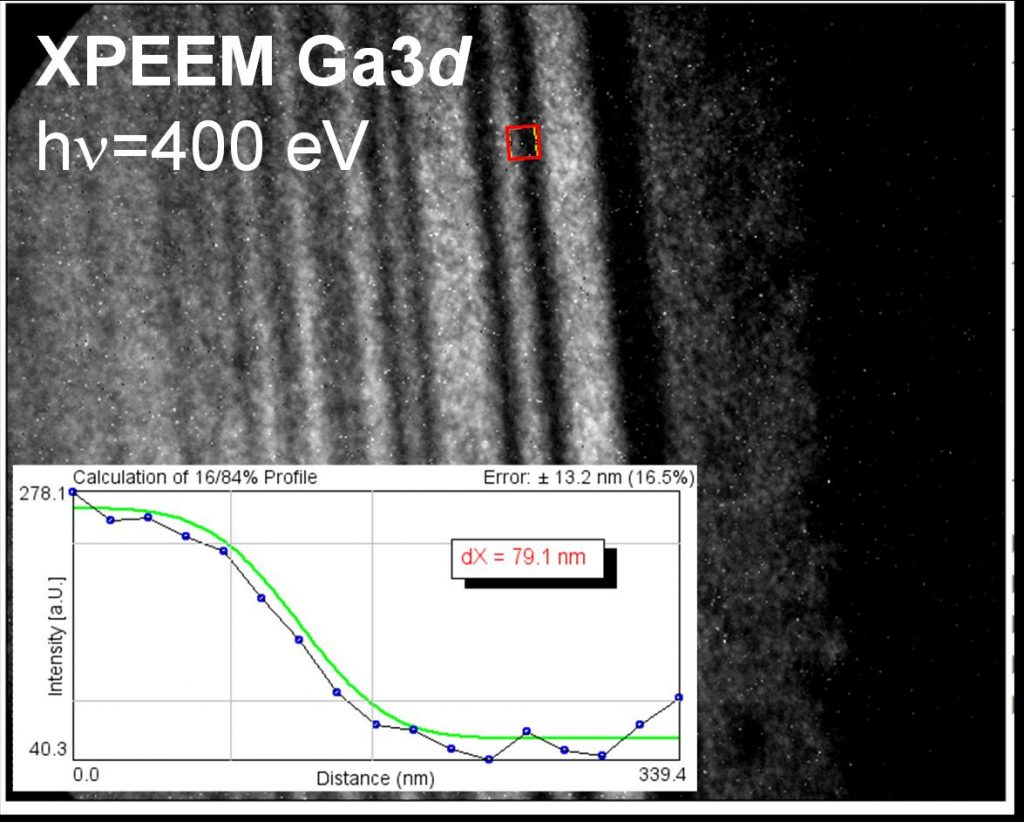
To characterize the instrument, energy filtered images of a standard sample certified of multi-layer GaAs/Ga0.3Al0.7As were realized at the Ga 3d core level (see figure). Contrast obtained following the Ga concentration gives a lateral resolution of 80 nm.
The very first experiments with the synchrotron radiation took place in February 2007. Among the studied systems and measurements appear:
• measurement of the chemical composition of the surface of SrTiO3 grains,
• measurement of the work function and surface composition of a Si nanofil capped by a Au-Si catalyst,
• study of the preferential polymer grafting on N-doped micronic zones of a Si substrate,
• study of p-Man electrochemically grafted Au/Tio2/SiO2 samples ,
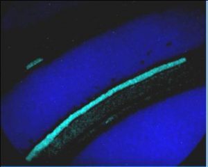
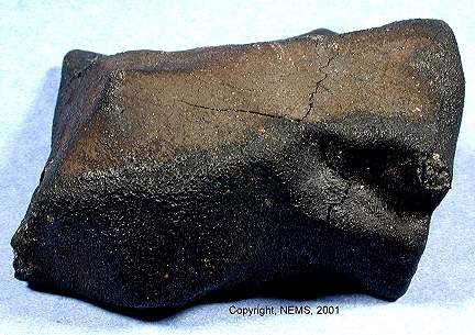
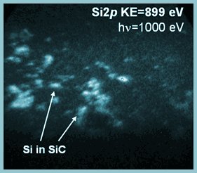
• in collaboration with the university of Mainz, identification, for the 1st time, of the pre-solar grains within the meteorite of Murchison from the localisation of SiC (Si(2p)) rich areas. NanoESCA seems to be the only instrument able to carry out this non-destructive chemical cartography much demanded by the astrophysicists in order to carry out isotopic ration analysis using nanoSIMS.
A new instrument with great possibilities for the surface physics!
Reference:
Applications of high lateral and energy resolution imaging XPS with a double hemispherical analyser based spectromicroscope,
M. Eschera, K. Winkler, O. Renault, N. Barrett, Journal of Electron Spectroscopy and Related Phenomena (2009).