Laëtitia Mayen, Nicholai D. Jensen, Maximilien Desbord, Danielle Laurencin, Christel Gervais, Christian Bonhomme, Mark E. Smith, Florence Porcher, Erik Elkaim, Cédric Charvillat, Pierre Gras, Christian Rey, Jérémy Soulié and Christèle Combes, CrystEngComm, 22 (2020) 3130-3143.
α-Canaphite (CaNa2P2O7·4H2O) is a layered calcium disodium pyrophosphate tetrahydrate phase of significant geological and potential biological interest. This study overcomes the lack of a reliable protocol to synthesize pure α-canaphite by using a novel simple and reproducible approach of double decomposition in solution at room temperature.
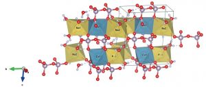
The pure α-canaphite is then characterized from the atomic to the macroscopic level using a multitool and multiscale advanced characterization strategy, providing for the first time full resolution of the α-canaphite monoclinic structure, including the hydrogen bonding network. Synchrotron X-ray diffraction and neutron diffraction are combined with multinuclear solid state NMR experimental data and computational modeling via DFT/GIPAW calculations. Among the main characteristics of the α-canaphite structure are some strong hydrogen bonds and one of the four water molecules showing a different coordination scheme. This peculiar water molecule could be the last to leave the collapsed structure on heating, leading eventually to anhydrous α-CaNa2P2O7 and could also be involved in the internal hydrolysis of pyrophosphate ions as it is the closest water molecule to the pyrophosphate ions.
Relating such detailed structural data on α-canaphite to its physico-chemical properties is of major interest considering the possible roles of canaphite for biomedical applications. The vibrational spectra of α-canaphite (deuterated or not) are analyzed and Raman spectroscopy appears to be a promising tool for the identification/diagnosis of such microcrystals in vitro, in vivo or ex vivo
https://doi.org/10.1039/D0CE00132E.


