


Pages scientifiques 2013

L’objectif principal de cette thèse consiste à développer une sonde à base de capteurs magnétorésistifs (Giant Magnetoresistance – GMR ou Tunnel Magnetoresistance - TMR) afin de mesurer le champ magnétique induit par les courants circulant le long des neurones. L’électrophysiologie classique permet actuellement d’enregistrer les potentiels électriques locaux des cellules nerveuses mais les électrodes utilisées n’apportent pas d’informations sur la signature magnétique de ces cellules. La sonde (appelée "magnétrode" en référence aux électrodes d’électrophysiologie) doit permettre d’obtenir la réponse électromagnétique des neurones, en utilisant la très bonne sensibilité et la possibilité de miniaturisation des capteurs magnétiques très sensibles (GMR ou TMR).
Dans un premier temps, la fabrication, la miniaturisation et la caractérisation des magnétrodes seront effectuées puis des tests in-vitro et in-vivo seront réalisés en collaboration avec respectivement l’équipe d’Alain Destexhe et Thierry Bal de l’Unité de Neurosciences, Information et Complexité (UNIC) du CNRS à Gif-sur-Yvette et celle du Professeur Pascal Fries de l’Ernst Strüngmann Institute (ESI) for Neuroscience in Cooperation with Max Planck Society à Francfort.
Titanium and its alloys are widely used in aeronautical, marine and chemical industries for their good mechanical properties, high resistance to corrosion, especially in sea water or human body, and low density, about 60% of steel density. However, tribological properties of titanium surface are poor with highly unstable friction coefficients and adhesive wear mechanisms which lead mechanical parts to seize up. Surface treatments are required to improve surface properties, according to the envisioned application.
Nitriding and carburizing are the most commonly used thermochemical treatments, but they must be done in highly oxygen-free atmospheres to avoid titanium oxidation. This rules out the possibility of local treatments. Laser induced modifications offer an alternative tool for modifying the surface composition by insertion of light elements (oxygen, nitrogen, carbon) present in the reactive atmosphere. Modified layers must have a high content of nitrogen to be hard enough to stand up to surface friction, while oxygen, localized in less crystallized phases, is also needed to accommodate friction without failure. Oxynitride formation appears then as ideal to reach optimal resistance.

Responsable : Sylvie MARGUET
Participants: Aurélie Habert, Jérôme Caron (Master-2), Mohammad Khaywah (Post-doc)
IRAMIS-NIMBE-LEDNA (Laboratoire Edifices Nanométriques)
Résumé: notre objectif est de tirer parti de l'interaction lumière-matière dans des nano-hybrides, constitués de nanoparticules d'or (AuNPs) colloïdales, dont la morphologie est optimisée, pour générer de la lumière, de la chaleur ou des porteurs de charge, selon l'application visée*.
* ce thème est en lien avec ceux développés au sein de deux groupements de recherche (GdR) du CNRS en France. Les GdR Or-nano et GdR PMSE (Plasmonique Moléculaire et Spectroscopies Exaltées).
… pour la PLASMONIQUE: depuis 2008 :
Collaborations : CEA-SPEC; L2n-Troyes; ISMO-Orsay; IS2M-Mulhouse; ILM-Lyon, ..
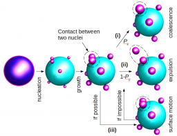
La plasmonique est une discipline à l'interface entre la physique, la chimie et la biologie. L’excitation de la résonance plasmon d’une nanoparticule (NP) métallique permet de générer des champs électromagnétiques très intenses, confinés à la surface de la NP, qui peuvent être utilisés pour exalter ou initier une réaction photochimique dans un « chromophore » placé à proximité. Le confinement du champ électromagnétique autour d’une NP colloïdale, monocristalline et non rugueuse, est supérieur à celui autour du même objet fabriqué par nanolithographie. La relaxation du plasmon, qui accompagne cet effet d’amplification, se fait selon des voies de relaxation ultra-rapides, en compétition les unes avec les autres. Leur importance relative dépend, de la morphologie de la NP, de son environnement "proche" et du mode d'irradiation. Ainsi, les NPs d’or peuvent se comporter comme des nanosources de lumière, de chaleur ou de porteurs de charges (électron, trou), selon la voie de désactivation qui prédomine. Pour ce qui est de la morphologie, c'est la taille, la présence de pointes et le rapport d'aspect (surface/volume) de la NP qui comptent. L’environnement désigne à la fois les «chromophores» adsorbés à la surface de la NP et notamment leurs niveaux d'énergie (HOMO-LUMO pour une molécule ou VB-CB pour un semi-conducteur) ainsi que la présence éventuelle d’une couche à l’interface (résidus de surfactant, couche isolante..). Enfin, le mode d'irradiation, continu ou pulsé à différentes échelles de temps de la micro- à la femto-seconde, est aussi un paramètre clé pour l’observation de ces trois types de nanosources.
Nous synthétisons des NPs d’or (Au-NP) en contrôlant minutieusement leur morphologie (taille, forme) et leur structure cristalline. Certaines de ces AuNPs ne sont produites que dans quelques laboratoires à travers le monde. Les microplaquettes (triangulaires, hexagonales ou en forme de disque), atomiquement planes, sont prometteuses pour la nanofabrication FIB de motifs monocristallins, non accessibles autrement. Un grand nombre de protocoles sont en cours d'élaboration pour des finalités diverses : i) remplacement du surfactant initial par d'autres molécules plus appropriées ; ii) dispersion homogène des NPs sur divers supports ; iii) auto-assemblage en une (1D), deux (2D), ou trois dimensions (3D) pour fabriquer des « points chauds » ; iv) enrobage par une couche de silice d'épaisseur variable. Des applications liées à l’environnement et à l’énergie sont envisagées, à plus long terme, via la synthèse de nanoparticules multimétalliques du type Au@X (avec X= Pd, Pt, Au, Ag or TiO2) qui combine un composant plasmonique (Au) et un catalyseur pour la catalyse plasmonique et la nanophotochimie induite par plasmon.
… pour la MEDECINE : depuis 2018 :
Collaborations: LPQM-CentraleSupelec, LAC-Orsay, CEA-SPEC, PPSM-ENS, ..
Nous produisons des nanoparticules cœur-coquille Au@silice qui ont un fort potentiel comme agent de contraste pour de nombreuses techniques d’imagerie médicale (photoacoustique, diffusion champ sombre, luminescence multi-photonique, Ultrasons à haute fréquence, contraste de phase quantitatif, tomographie par ordinateur…). En ce sens, le projet ANR SINAPSE, récemment accepté vise à développer une nouvelle classe de NP pour l'imagerie neuronale (coord F. Marquier). Les NPs d’or sont d’excellents capteurs (e.g test de grossesse) du type LSPR, SERS, Fluorescence, Pression ... Dans le domaine de la photothérapie, les NPs d'or présentant un rapport d'aspect (longueur/épaisseur) élevé, tels que les fils et les plaquettes, sont très intéressantes car elles peuvent être excitées par des photons infrarouges, dans les premières fenêtres (I,II,III,IV) de transparence des tissus biologiques. La génération de chaleur induite par plasmon (PTT ; photothermal therapy) et plus récemment la génération de R.O.S. (reactive oxygen species) à partir de NPs d’or seules (c.a.d. sans photosensiblisateur) est une approche originale pour traiter les tumeurs, en particulier en absence d'oxygène, lorsque la PDT (photodynamic therapy) conventionnelle, basée sur l’utilisation de molécules type porphyrine à l'état triplet excité triplet, ne peut pas fonctionner. Le projet HEPPROS (plan cancer) a pour but d’étudier la génération de chaleur et de R.O.S. telles que 1O2, OH., O2.-, H2O2 pour diverses morphologies de NPs d’or et ainsi mieux comprendre les mécanismes transitoires complexes impliqués à l'interface AuNP/adsorbat moléculaire (coord B. Palpant).
Heterogeneous samples are usually studied using solid-state NMR methods and while the sample is spinning at the magic angle. This is problematic for many cases, where the sample cannot be spun, as in MRI. The alternative methodology of spinning the magnetic field around a static sample has been proposed since the 60's, but not yet successfully applied. Our goal is to develop the methodology and the instrumentation in order to perform magic angle field spinning and answer questions related to the monitoring of metabolism in living matter as well as the structure and function of complex systems like batteries, porous media and fuel cells.
Portable, high-sensitivity low-field detectors are the second axis of our research. Analysis away from the laboratory becomes important, in particular in hostile environments, such as high-radiation areas. It can be also cost effective compared to standard superconducting imaging systems. Imaging and on-line inspection of such industrial processes is a challenging question we try to solve.
There are many cases where the sample, object or subject we want to study cannot be brought at the laboratory and be placed inside the bore of a high field super-conducting magnet. Such situations appear often in industrial and biomedical applications. Additionally in many case access is limited to one half of the space (for example when trying to measure moisture inside a wall), and a one-sided detection system is needed. This direction of research was initiated with the project named NMR2GO funded by ANR and has now reached the point were uniform one-sided magnets are built and are operational. The goal of the project is to design and built uniform permanent magnets with MRI characteristics. One such magnet is shown on the figure on the left. It generates a uniform gradient above its surface, which remains linear for many cm inside the object under study. Applications of this device in surface sciences are currently underway.
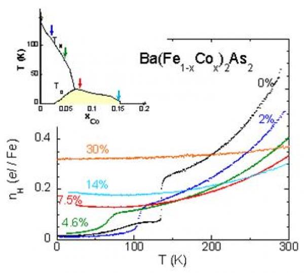
The multiband nature of the iron-based superconductors is a key factor to understanding their physical properties. We have performed a complete study of the charge transport in Ba(Fe1-xCox)2As2 single crystals, in connection with a determination of their electronic structures by photoemission experiments.
In the new Fe-based pnictide superconductors discovered in 2008, the appearance of high-Tc superconductivity in close proximity to the antiferromagnetic phase has been taken as the signature of unconventional superconductivity. It seems now well established that magnetism and superconductivity are directly connected to the peculiar features of the electronic structures of these compounds, which are characterized by small hole and electron pockets. The transport properties of the prototypical Ba(Fe1-xCox)2As2 have been studied throughout the electron doped phase diagram as fine tuning of Co content can be achieved in sizeable single crystals.

The UV-induced DNA lesions at mutational hotspots are not randomly distributed but depend on the base sequence around them. This effect could be related to the redistribution of the electronic excitation energy among the bases. During the past term, we had evidenced that ultrafast energy transfer takes place in model duplexes with repetitive base sequence; this process is possible because the initially populated states are delocalized on more than one base (excitons). It was predicted that this phenomenon should not occur in natural DNA since it is a highly disordered system. Studying the fluorescence of natural DNA, we showed that this prediction was not correct. In this case, the energy transfer process can be mediated by interconversion between ππ* states and charge transfer states. As a result ππ* excitations exhibit multiscale decay, from the femtosecond to the nanosecond time-scale.
The figure shows how photon absorption populates exciton states which may be trapped by charge transfer states. Charge separation and charge recombination to ππ* states gives rise to delayed fluorescence (bold green). Minor contributions to ππ* fluorescence arise during intraband scattering and/or localization of the excitons (thin green).
Natural DNA publications:
I. Vaya, T. Gustavsson, F.A. Miannay, T. Douki, D. Markovitsi: Fluorescence of Natural DNA: From the Femtosecond to the Nanosecond Time Scales, J. Am. Chem. Soc., 132: 11834. 2010.
I. Vayá, T. Gustavsson, T. Douki, Y. Berlin, D. Markovitsi: Electronic Excitation Energy Transfer between Nucleobases of Natural DNA, J. Am. Chem. Soc., 134: 11366. 2012.
P. Changenet-Barret, T. Gustavsson, D. Markovitsi, I. Manet
ChemPhysChem 2016.
Drug/Protein Interactions Studied by Time-Resolved Fluorescence Spectroscopy
T. Gustavsson, D. Markovitsi, I. Vaya, P. Bonancia, M. Consuelo Jimenez, M. A. Miranda
Physical Chemistry of Interfaces and Nanomaterials Xiii 2014, 9165.
I. Vayá, P. Bonancía, M. C. Jiménez, D. Markovitsi, T. Gustavsson, M. A. Miranda
Phys. Chem. Chem. Phys. 2013, 116, 4727-4734
P. Changenet-Barret, T. Gustavsson, D. Markovitsi, I. Manet, S. Monti
Phys. Chem. Chem. Phys. 2013, 15, 2937-2944.
P. Bonancía, I. Vayá, D. Markovitsi, T. Gustavsson, M. Consuelo Jiménez, M. A. Miranda
Org. Biomol. Chem. 2013, 11, 1958-1963.
Excited-State Interactions in Diastereomeric Flurbiprofen–Thymine Dyads
P. Bonancía, I. Vayá, M. J. Climent, T. Gustavsson, M. Markovitsi, C. Jiménez, M. A. Miranda
J. Phys. Chem. A 2012, 116, 8807−8814.
Femtosecond Spectroscopic Study of Carminic Acid–DNA Interactions
R. Comanici, B. Gabel, T. Gustavsson, D. Markovitsi, C. Cornaggia, S. Pommeret, C. Rusu, C. Kryschi
Chem. Phys. 2006, 325, 509-518.
-
ANR-17-CE29-0008 TUNIFOLD-S Tuning foldable building blocks using sulfur; 2018-2020; Coord. Michel Mons; Partenaires D.J. Aitken (ICMMO, Orsay) et Anne Zehnacker (ISMO, Orsay)
-
ANR-16-CE29-0017 IONPAIRS Spectroscopy of isolated and microsolvated ion pairs; 2016-2020; Coord. E. Gloaguen
-
ANR-14-CE06-0019 ESBODYR Excited States of BiO-relevant systems: towards ultrafast DYnamics with conformational Resolution; 2014-2018; Coord. V. Brenner, Part. S. Hoyau (Toulouse), A. Zehnacker, ISMO (Orsay) - site du projet ESBODYR
-
Projet LabEx PALM 2013 "DIRCOS": IR Diagnostic IR for a COnformation-Selective femtochemistry; Coord. L. Poisson, LFP/DyR, Partenaires E. Gloaguen, LFP/SBM; A. Zehnacker, ISMO (Orsay), P. D'Oliveira, SPAM/SLIC
-
DRF Impulsion Invention, projet CompaTREG 2018 (port. LIDYL M. Géléoc; partenaires J-P Renault /NIMBE et T. Oksenhendler /iTEOX)
-
-
PALM Valo, projet SEIEM 2019 (port. LIDYL M. Géléoc ; partenaire J-P Renault /NIMBE),
-
DIM Analytics, projet EPuR 2017-2020 (port. LIDYL M. Géléoc), partenaires J-P Renault (NIMBE) et T. Oksenhendler (iTEOX)
-
Accès aux moyens de calcul des centres de calcul nationaux ( CCRT/CINES/IDRIS) au travers de l'allocation de ressources attribuée par GENCI (Grand Equipement National de Calcul Intensif) pour nos travaux théoriques
|
Permanents · Dr. François PIUZZI Retired in 2013 · Dr. Iliana DIMICOLI Retired in 2006 · Dr. Rodolphe POLLET 2008-2010
Post-docs · Dr. Thibaut VERY 2015-2016 · Dr. Woon-Yong SOHN 2015-2016 · Dr. Hiroya ASAMI 2014-2015 · Dr. Vanesa VAQUERO-VARA 2014-2015 · Dr. Mohammad ALAUDIN 2013-2014 · Dr. Manuela CIRTOG 2011-2012 · Dr. Richard J. PLOWRIGHT 2010-2011 · Dr. Himansu S. BISWAL 2009-2011 · Dr. Francesca PAGLIARULO 2006-2007
Doctorants · Sana HABKA 2014-2017 · Yohan LOQUAIS 2010-2013
Visiteurs · Dr. Nadia DOSLIC (IRB Zagreb, Croatie) Août 2012 · Momir MALIS (IRB Zagreb, Croatie) Nov-Dec 2012 · Dr. Jessica THOMAS (Pittsburgh, USA) Jan-Avr 2008 · Prof. E.R. BERNSTEIN (Fort-Collins, CO, USA) Avr. Mai 2008 · Dr. Marco AROLD (Iena, Allemagne) Nov-Dec 2008 · Prof. Hiroyuki SAIGUSA (Yokohama, Japon) Fev-Mar 2007 |
Epitaxial graphene grown on SiC substrate has been intensively studied over the past ten years, in view of its applications in nanoelectronics. The C-terminated face of the substrate offers a particular interest due to its rotational stacking, which decouples adjacent graphene sheets. A major issue is the microscopic surface heterogeneity giving, for example, domains with varying numbers of graphene layers with different electronic and chemical properties. Energy-filtered photoelectron emission microscopy (PEEM) can correlate real space chemical and work function mapping with reciprocal space imaging of the complete band structure of micron scale regions. Therefore, this technique allows a better understanding of the local properties – morphology, chemistry of the interface and electronic properties – which is still required for the development of new electronic devices.
Bright field real and reciprocal space imaging
The photoemission threshold is directly related to the work function (WF) of a material. By fitting the spectrum of each pixel of an image series, acquired over the photoemission threshold, it is therefore possible to map the work function value over the field of view (FoV) of the microscope. This procedure has been applied on an image series of epitaxial graphene on SiC(000-1) (figure 1a). The work function variation is thought to be due to the graphene thickness and the chemistry of the graphene/SiC interface. Similarly, the intensity map of the SiC component of C 1s core level can be used to estimate the thickness variation of the graphene, and can be correlated to the WF map. When the number of graphene layer increases, the SiC peak intensity decreases, relative to the graphene one. In this way, the local thickness of the graphene film can be estimated from the relative SiC and graphene C 1s intensity; within the FoV graphene domains between 1 and 3 monolayers thick are identified.
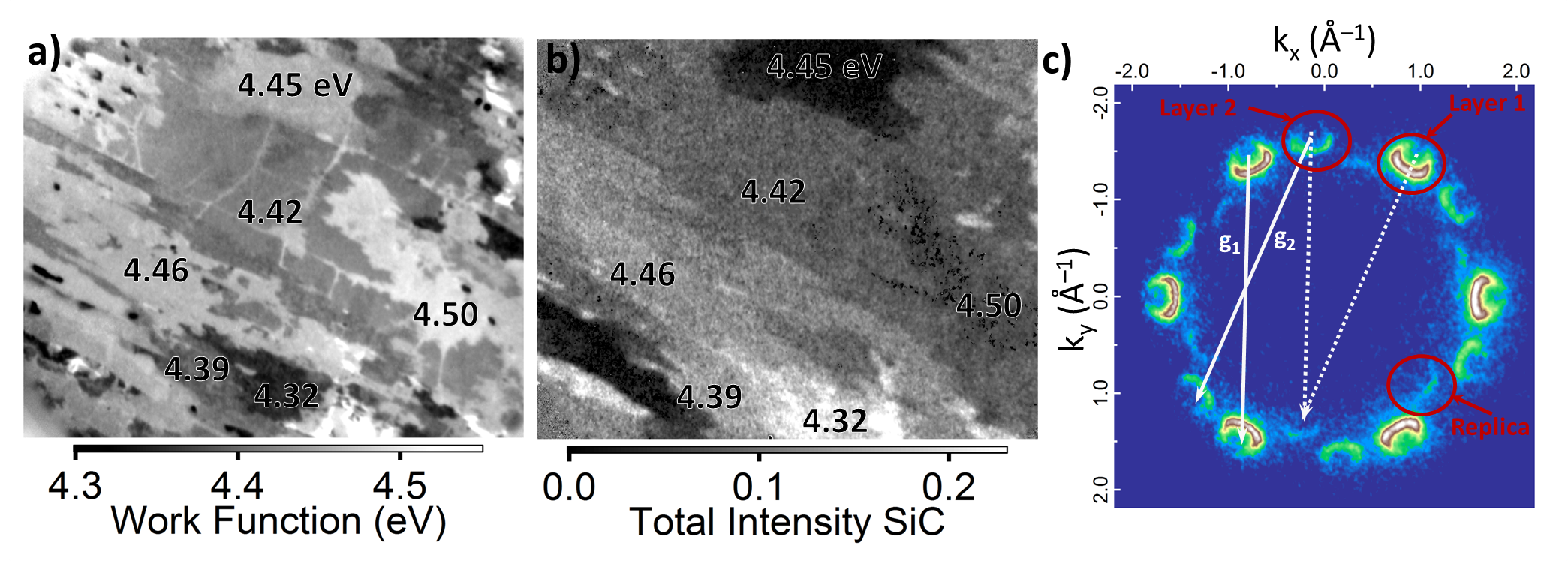
Figure 1: a) Work function map of a 53 μm FoV, using a photon energy of 654.3 eV. b) Intensity map (arbitrary units) of the SiC substrate component of C 1s spectrum after background subtraction for a FoV of 34 μm. c) Constant energy cut in the k-space below the Dirac point, acquired at a binding energy of 1.3 eV, for a graphene bilayer area.
Thanks to the easy switching between the real space and the reciprocal space imaging modes of the NanoESCA (Omicron Nanotechnology), it is possible to obtain the complete band structure of a given area down to 10 µm2, by inserting a field aperture (FA) to select the desired region. A constant energy cut of a graphene bilayer in the k-space below the Dirac point is presented in figure 1c. It shows the presence of two sets of Dirac cones, with the well-known sixfold symmetry. They are attributed to two commensurately rotated graphene sheets, with a rotation angle of 21.9°. A fainter feature presenting also a sixfold symmetry, which appears inside the radius of the most intense ones, is due to a diffraction resulting from the supercell (g1 and g2) formed by the commensurate rotations, as shown in the figure 1c. The reciprocal space imaging mode of the NanoESCA allows immediate visualization of these diffracted cones because it probes all wave vectors parallel to the sample surface.
Dark field imaging
Dark field imaging has been performed using focused He lamp (He I =21.2 eV), on such epitaxial graphene sample grown on a SiC(000-1) substrate. The real space image of the FoV at the photoemission threshold is shown in figure 2a. Figure 2b presents a constant energy cut of the band structure, averaged over the whole FoV. Six commensurate rotation angles are observed (a-f). The contrast aperture in the diffraction plane of the microscope has then been closed to a diameter of 0.11 Å-1 at each of the Dirac cones and the microscope has been switched back to the real space mode. The corresponding real space image shows the spatial origin of photoelectrons for a specific emission angle. By repeating this experiment for each rotational angle, a spatial distribution of the commensurate rotations has been determined (figure 2c). The Dirac cones at the K and K’ point of the first Brillouin zone, typical of graphene, can therefore be used to determine a real spatially-resolved map as function of the photoelectron wave vector.

Figure 2: a) Real space image of the FoV at the photoemission threshold. b) Constant energy cut in the k-space below the Dirac point showing the commensurately rotations of the graphene sheets. c) Reconstructed valence band image from the sum of the dark field images, acquired for the six commensurate rotation angles.
The PEEM study of epitaxial graphene on SiC(000-1) allows the correlation of spatial imaging, which links the WF variation to the number of graphene layer along with its interface chemistry, and to the local band structure, highlighting diffraction effects due to commensurate rotations of the graphene sheets. Dark field PEEM completes the information with a spatially resolved map of the commensurately rotated graphene sheets.
For more information, read the following paper:
Microscopic correlation between chemical and electronic states in epitaxial graphene on SiC(000-1), C. Mathieu, N. Barrett, J. Rault, Y. Mi, B. Zhang, W. A. de Heer, C. Berger, E. Conrad and O. Renault, Physical Review B 83, 235436 (2011).
Laboratory-based real and reciprocal space imaging of the electronic structure of few layer graphene on SiC(000-1) using photoelectron emission microscopy, N. Barrett, K.Winkler, B.Kromker, E.H.Conrad, Ultramicroscopy 130, 94 (2013).
Microscopic chemical and electronic structure of few layer graphene on SiC(000-1), C. Mathieu and N. Barrett, PICO, The Omicron Nanoscience Newsletter 17, 6-8 (2013)
« Back to the Group page « Back to the Oxides page
Domain walls are intrinsic to ferroic materials. Long considered as understood, it is only recently, thanks to atomic-resolution studies using transmission electron microscopy (TEM) or atomic force microscopies (AFM, c-AFM, PFM etc.) that their real structural and electronic complexity has been revealed. Based on this, a new paradigm of ferroic devices is now envisaged where the domain walls, rather than the domains, are the active element. This field has been coined “Domain boundary engineering” or “Domain wall nanoelectronics”.
Photoferroelectrics are materials with both photoelectric and ferroelectric properties. As correlated-electron materials, they are attracting increasing interest because of their high technological potential. Recent reports show that low conversion efficiencies can be counterbalanced by large, above-band gap photo-voltages in complex multiferroic oxides and the fundamental role of the potential difference across domain walls.
More detailed and quantitative knowledge of its structural, electronic and chemical nature is necessary. Fundamental questions on the crystal structure and the electronic structure in the vicinity of the domain wall, as well as their behavior under illumination remain to be elucidated.
« Back to the Group page « Back to the Oxides page
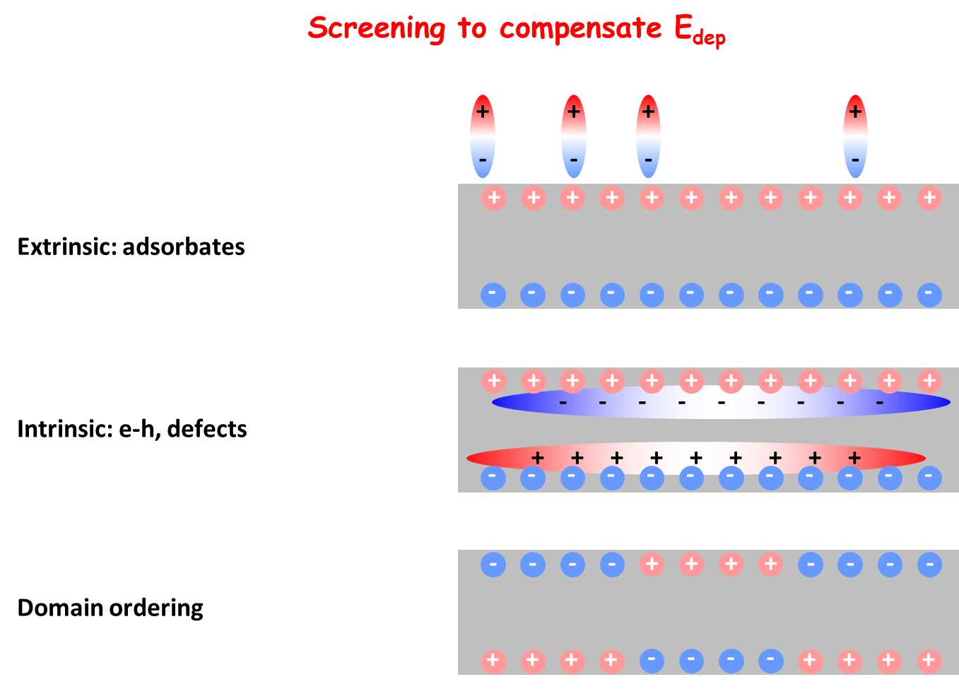
Switching requires a metallic contact, raising fundamental issues about the interface between the FE and the electrode. Polarization leads to fixed charge of opposite sign at the two metal-FE interfaces. Electrode free charge acts to screen the polarization charge creating dipoles of the same sign at the two interfaces, however, the screening is always imperfect and the residual depolarizing field alters the electrostatic potential inside the FE. The partially covalent nature of the bonds in the FE changes the band structure with respect to that of a perfectly ionic compound which, from consideration of dynamical charge tensors, depends on the atomic distortion. Reduction/oxidation conditions at the surface can control the film polarization.
« Back to the Group page « Back to the Oxides page
Surface polarization charge in ferroelectric (FE) materials can be screened by a variety of mechanisms: intrinsic (charge carriers or defects in the bulk), extrinsic (chemical environment or adsorbates), domain ordering, or even a combination of the above. Chemisorption of OH- and protons can lead to important changes in the electrical boundary conditions and water film can play an active role in domain switching. Photo-generated charge carriers can also efficiently screen the surface and interface polarization charge and hence the depolarizing field. The photo-generated pair lifetimes depend on the FE surface topology and the polarization state can influence, for example, redox reactions
Alexandre Hervé, Pierre Thuéry, Michel Ephritikhine, Jean-Claude Berthet
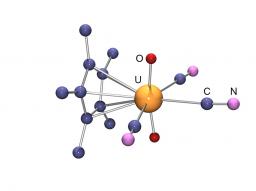
L’ion cyanure CN− est certainement l'un des ligands les plus utilisés en chimie de coordination des métaux de transition d. De nombreuses études ont montré l’intérêt des complexes cyanures dans des domaines très variés allant de la biologie aux nanomatériaux. En plus d’une riche chimie de coordination liée aux divers modes de coordination du group CN, les complexes cyanures présentent bien souvent de remarquables propriétés magnétiques, photoluminescentes et/ou de conductivité. Au contraire de celle des métaux d, la chimie des cyanures des métaux f (lanthanides et actinides) n’a été que très peu considérée.
Le laboratoire LCCEf s’intéresse depuis quelques années au développement de cette thématique à partir de précurseurs organométalliques ou comportant des ligands azotés, et a mis en évidence le potentiel du ligand CN−,
- pour stabiliser les nombreux degrés d’oxydation de l’uranium1-4
- et former des espèces inattendues (métallocènes linéaires,2,3 complexe cyclopentadiènylique de l’uranyle4) bouleversant ainsi les mentalités et ouvrant de nouvelles possibilités notamment en chimie organométallique.
Les études actuelles concernent la formation de briques moléculaires mono ou polycyanures, intéressantes pour de nouveaux développements en chimie de coordination, mais surtout susceptibles de conduire à la formation de clusters et d’agrégats homo ou polymétalliques originaux dont les propriétés photochimiques (magnétisme et luminescence) seront étudiées. Ces études expérimentales couplées à des calculs théoriques favoriseront également une compréhension plus fine de la liaison Actinide-ligand nécessaire sur les plans fondamental et appliqué (Nucléaire).
The parahydrogen induced polarization (PHIP) method is based on the non-Boltzmann distribution of nuclear spin states following the hydrogenation of a substrate by parahydrogen. Absorptive and/or emissive NMR signals can be greatly enhanced, up to several orders of magnitude as compared to thermally polarized molecules.
The research in this field is part of the expertise of LSDRM in its effort to increase NMR sensitivity.
Dihydrogen, initially made of 1/4 of parahydrogen and 3/4 of orthohydrogen, a proportion that does not induce any hyperpolarization, is cooled to -196°C in the presence of a ferric catalyst, thanks to an home-made apparatus. The resulting gas contains about one half of parahydrogen, and may be use to obtain hyperpolarized spectra.
A first example: the method named "signal amplification by reversible-exchange" (SABRE) is used here to increase the aromatic signals of N,N-dimethylnicotinamide. In SABRE, a catalyst reversibly binds dihydrogen and the aromatic molecule.
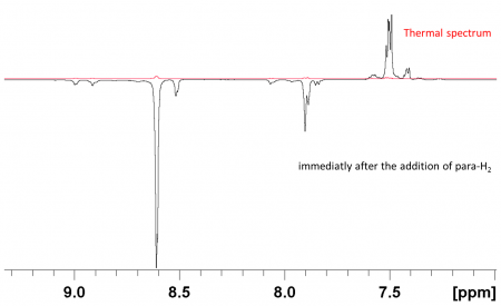
Le CFC (Carbon Fiber Composite) est utilisé depuis longtemps comme matériau de première paroi dans les tokamaks pour la fusion thermonucléaire. Il présente de nombreux avantages du fait de ses propriétés thermomécaniques remarquables et son faible numéro atomique, contribuant peu au refroidissement du plasma par rayonnement. L’inconvénient est qu’il possède une capacité forte à piéger le combustible (dans le cas des tokamaks d’étude, le deutérium). L’utiliser dans un cadre de production d’énergie, avec du tritium, pose des problèmes de sureté du fait des limites imposées en termes d’inventaire.
PhotoEmission
MesoXcope
XPD
LEEM
Common characterization techniques
We use time-resolved fluorescence spectroscopy from the femtosecond to the nanosecond time scales to study the dynamics of the interaction between various types of drugs (anti-inflammatory, antitumor, antidiabetic..) and biomolecules (DNA, G-quadruplexes, aninoacids, glycogen…).
These studies are performed in the frame of SLIC/LaserLab Europe in collaboration with colleagues from Germany, Spain, Italy and Greece.
Ce dispositif expérimental est doté d’un jet versatile couplé à un dispositif d’imagerie de photoélectron et à un spectre de masse à temps de vol. L’objectif de cette expérience est de deux ordres :
- L’étude de la spectroscopie des états de transition de complexes de van der waals. Les réactions métaux-molécules ont été particulièrement étudiées.
- La détermination de la spectroscopie d’absorption de différents systèmes.
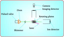
Responsable : Marc Briant/Marc-André Gaveau
Cette équipe étudie les mécanismes par lesquels la solvatation affecte la dynamique d’une réaction chimique. Le travail a porté jusqu'à présent sur des réactions d'espèces excitées électroniquement. Une évolution est en cours vers les réactions qui se déroulent à l’état électronique fondamental mais où l'excitation vibrationnelle joue un rôle déterminant.
Responsables : Christian Angelié / Jean-Maïk Soudan
Cette équipe a pour ambition d'apporter une assise numérique réaliste à des concepts aussi importants que changement de phase, ségrégation chimique et d’auto-organisation dans les systèmes Fer/carbone. Des algorithmes et des "codes maisons" sont développés pour traiter de ces questions sur des agrégats comportant plusieurs centaines, voire plusieurs millier d'atomes. Il y a deux aspects à ce travail :
- Développement algorithmique de type Monte Carlo pour aborder des questions de thermodynamique
- Développements méthodologiques sur des potentiels empiriques de type réactif, avec inclusion de propriété magnétiques, pour traiter de l'interaction du fer avec un environnement
Cet axe de recherche a deux motivations:
- Développer de nouveau concepts à même de faire progresser les techniques de simulation numérique des gros systèmes;
- Apporter des outils permettant de modéliser la croissance de nanotubes de carbone sur un catalyseur de fer.
Cet axe a été porté en partie par les programmes nationaux CHIMTRONIC et NANOSIMUL du CEA.
Bibliographie
- J. M. Soudan, M. Basire, J. M. Mestdagh et C. Angelie, A new Monte Carlo method for getting the density of states of atomic cluster systems, Journal of Chemical Physics 135, 144109 (2011) & Erratum Journal of Chemical Physics 135, 229901 (2011)
- M. Basire, J. M. Soudan et C. Angelie, Nanothermodynamics of large iron clusters by means of a flat histogram Monte Carlo method, Journal of Chemical Physics 141, 104304 (2014)
Epitaxial graphene grown on SiC substrate has been intensively studied over the past ten years, in view of its applications in nanoelectronics. The C-terminated face of the substrate offers a particular interest due to its rotational stacking, which decouples adjacent graphene sheets. A major issue is the microscopic surface heterogeneity giving, for example, domains with varying numbers of graphene layers with different electronic and chemical properties. Energy-filtered photoelectron emission microscopy (PEEM) can correlate real space chemical and work function mapping with reciprocal space imaging of the complete band structure of micron scale regions. Therefore, this technique allows a better understanding of the local properties – morphology, chemistry of the interface and electronic properties – which is still required for the development of new electronic devices.
Bright field real and reciprocal space imaging
The photoemission threshold is directly related to the work function (WF) of a material. By fitting the spectrum of each pixel of an image series, acquired over the photoemission threshold, it is therefore possible to map the work function value over the field of view (FoV) of the microscope. This procedure has been applied on an image series of epitaxial graphene on SiC(000-1) (figure 1a). The work function variation is thought to be due to the graphene thickness and the chemistry of the graphene/SiC interface. Similarly, the intensity map of the SiC component of C 1s core level can be used to estimate the thickness variation of the graphene, and can be correlated to the WF map. When the number of graphene layer increases, the SiC peak intensity decreases, relative to the graphene one. In this way, the local thickness of the graphene film can be estimated from the relative SiC and graphene C 1s intensity; within the FoV graphene domains between 1 and 3 monolayers thick are identified.
Figure 1: a) Work function map of a 53 μm FoV, using a photon energy of 654.3 eV. b) Intensity map (arbitrary units) of the SiC substrate component of C 1s spectrum after background subtraction for a FoV of 34 μm. c) Constant energy cut in the k-space below the Dirac point, acquired at a binding energy of 1.3 eV, for a graphene bilayer area.
Thanks to the easy switching between the real space and the reciprocal space imaging modes of the NanoESCA (Omicron Nanotechnology), it is possible to obtain the complete band structure of a given area down to 10 µm2, by inserting a field aperture (FA) to select the desired region. A constant energy cut of a graphene bilayer in the k-space below the Dirac point is presented in figure 1c. It shows the presence of two sets of Dirac cones, with the well-known sixfold symmetry. They are attributed to two commensurately rotated graphene sheets, with a rotation angle of 21.9°. A fainter feature presenting also a sixfold symmetry, which appears inside the radius of the most intense ones, is due to a diffraction resulting from the supercell (g1 and g2) formed by the commensurate rotations, as shown in the figure 1c. The reciprocal space imaging mode of the NanoESCA allows immediate visualization of these diffracted cones because it probes all wave vectors parallel to the sample surface.
Dark field imaging
Dark field imaging has been performed using focused He lamp (He I =21.2 eV), on such epitaxial graphene sample grown on a SiC(000-1) substrate. The real space image of the FoV at the photoemission threshold is shown in figure 2a. Figure 2b presents a constant energy cut of the band structure, averaged over the whole FoV. Six commensurate rotation angles are observed (a-f). The contrast aperture in the diffraction plane of the microscope has then been closed to a diameter of 0.11 Å-1 at each of the Dirac cones and the microscope has been switched back to the real space mode. The corresponding real space image shows the spatial origin of photoelectrons for a specific emission angle. By repeating this experiment for each rotational angle, a spatial distribution of the commensurate rotations has been determined (figure 2c). The Dirac cones at the K and K’ point of the first Brillouin zone, typical of graphene, can therefore be used to determine a real spatially-resolved map as function of the photoelectron wave vector.
Figure 2: a) Real space image of the FoV at the photoemission threshold. b) Constant energy cut in the k-space below the Dirac point showing the commensurately rotations of the graphene sheets. c) Reconstructed valence band image from the sum of the dark field images, acquired for the six commensurate rotation angles.
The PEEM study of epitaxial graphene on SiC(000-1) allows the correlation of spatial imaging, which links the WF variation to the number of graphene layer along with its interface chemistry, and to the local band structure, highlighting diffraction effects due to commensurate rotations of the graphene sheets. Dark field PEEM completes the information with a spatially resolved map of the commensurately rotated graphene sheets.
For more information, read the following paper:
Microscopic correlation between chemical and electronic states in epitaxial graphene on SiC(000-1), C. Mathieu, N. Barrett, J. Rault, Y. Mi, B. Zhang, W. A. de Heer, C. Berger, E. Conrad and O. Renault, Physical Review B 83, 235436 (2011).
Laboratory-based real and reciprocal space imaging of the electronic structure of few layer graphene on SiC(000-1) using photoelectron emission microscopy, N. Barrett, K.Winkler, B.Kromker, E.H.Conrad, Ultramicroscopy 130, 94 (2013).
Microscopic chemical and electronic structure of few layer graphene on SiC(000-1), C. Mathieu and N. Barrett, PICO, The Omicron Nanoscience Newsletter 17, 6-8 (2013)
« Back to the Group page « Back to the Oxides page
Switching requires a metallic contact, raising fundamental issues about the interface between the FE and the electrode. Polarization leads to fixed charge of opposite sign at the two metal-FE interfaces. Electrode free charge acts to screen the polarization charge creating dipoles of the same sign at the two interfaces, however, the screening is always imperfect and the residual depolarizing field alters the electrostatic potential inside the FE. The partially covalent nature of the bonds in the FE changes the band structure with respect to that of a perfectly ionic compound which, from consideration of dynamical charge tensors, depends on the atomic distortion. Reduction/oxidation conditions at the surface can control the film polarization.
« Back to the Group page « Back to the Oxides page
Surface polarization charge in ferroelectric (FE) materials can be screened by a variety of mechanisms: intrinsic (charge carriers or defects in the bulk), extrinsic (chemical environment or adsorbates), domain ordering, or even a combination of the above. Chemisorption of OH- and protons can lead to important changes in the electrical boundary conditions and water film can play an active role in domain switching. Photo-generated charge carriers can also efficiently screen the surface and interface polarization charge and hence the depolarizing field. The photo-generated pair lifetimes depend on the FE surface topology and the polarization state can influence, for example, redox reactions
Parmi les différents paramètres impliqués dans les mécanismes d'accélération laser-plasma, les électrons eux-mêmes peuvent être détectés et étudiées. Mais, comme ils sont détecter à la fin du processus d'accélération, il n'est pas trivial d’obtenir des informations sur la «vie de l 'électron", de l’injection jusqu’à la fin du processus d’accélération. Pendant la phase d'accélération, les électrons acquièrent des énergies relativistes et oscillent autour de l'axe du laser. Cela conduit à la production d'émission, appelé rayonnement betatron, jusqu'aux rayons g [Cipiccia2011 ]. Plusieurs études expérimentales et numériques ont montré qu'il existe un lien étroit entre les propriétés électroniques et les caractéristiques d'émission bêtatroniques [Rousse2007, Ta Phuoc2006].
Nous avons effectué des études numériques sur les propriétés de l'émission betatron des électrons relativistes [ Andre2012 ] dans des conditions proches de celles pouvant être obtenues sur le laser UHI100 (CEA-SLIC100TW-25fs).
D'après les simulations 2D PIC que nous avons effectuées avec PICLS (collaboration avec E. d'Humières - CELIA-Université Bordeaux1) couplé à un programme développé par Arnaud André (doctorant) pour calculer l’émission des électrons accélérés, nous avons montré que les propriétés du rayonnement betatron sont fortement liés à ceux du faisceau d'électrons. De légères variations des conditions initiales (figure 1), la densité électronique variant de 6 à 7x1018cm-3, permettent de lier les propriétés du paquet d'électrons à la fin du processus d'accélération (phase d'oscillation, de l'énergie, nombre d'électrons dans chaque grappe, lorsque plusieurs paquets d'électrons piégés) au profil du rayonnement émis et à l'étendue spectrale de la répartition angulaire sur l'axe de propagation du laser
[André2012].
ELISA (ELectron Injector for compact Staged high energy Accelerator) : un injecteur d’électrons pour un schéma d’accélération multi-étages (collaborations : LPGP-Université Paris Sud / LULI-Ecole Polytechnique).
 Le projet ELISA consiste à mettre au point expérimentalement une source contrôlée d’électrons monocinétiques, destinée à servir d’injecteur pour un accélérateur laser-plasma multi-étages (Fig. 1). Disposer d’un injecteur approprié et fiable est une nécessité pour l’accélération à plusieurs étages, qui semble être la voie la plus prometteuse pour produire des électrons à des énergies ultra-relativistes de façon compacte et contrôlée. La production d’électrons d’énergie accordable entre 10 et 100 MeV sera étudiée dans une cellule de longueur variable, remplie d’un mélange de gaz permettant de contrôler le piégeage d’électrons par ionisation. Des simulations numériques seront réalisées pour optimiser l’injecteur en fonction des paramètres du laser puis pour décrire le couplage et l’accélération du faisceau d’électrons obtenu, dans un étage accélérateur de grande longueur (>10 cm).
Nous avons effectué des simulations numériques avec le 2D PIC Code Calder (coll. Erik Lefebvre/Rachel Nuter - CEA-DAM BIII) pour comprendre l'évolution temporelle de l'impulsion laser ultra-courte et à haute intensité dans le plasma. Tout d'abord, nous avons testé le code en simulant la propagation du laser dans les conditions d’une expérience réalisée par les collaborateurs de l'Université de Pise et avons pu décrire et interpréter une partie de leurs résultats expérimentaux. Il s’agissait d’étudier l'évolution temporelle de l'impulsion laser dans un plasma généré avec une intensité laser de 3.5x10¹⁸W/cm2 et Ne0 = 1019cm-3 (azote et l'argon plasma) [Giulietti 2013]. Nous avons isolé différentes contributions dominantes du plasma lors de la propagation, sur le spectre de l'impulsion laser (décalage vers le bleu et le rouge de l'impulsion laser en raison de l’auto-modulation par l'onde de plasma - décalage vers le bleu par le gradient de densité électronique décroissant).
Enfin, nous avons examiné les mêmes paramètres d'entrée («régime de la bulle" accessible avec UHI100laser - 100TW-25fs) que ceux correspondant à notre étude de l'émission betatron et nous avons décrit l'évolution temporelle de la propagation des impulsions laser dans le plasma.
The UV-induced DNA lesions at mutational hotspots are not randomly distributed but depend on the base sequence around them. This effect could be related to the redistribution of the electronic excitation energy among the bases. During the past term, we had evidenced that ultrafast energy transfer takes place in model duplexes with repetitive base sequence; this process is possible because the initially populated states are delocalized on more than one base (excitons). It was predicted that this phenomenon should not occur in natural DNA since it is a highly disordered system. Studying the fluorescence of natural DNA, we showed that this prediction was not correct. In this case, the energy transfer process can be mediated by interconversion between ππ* states and charge transfer states. As a result ππ* excitations exhibit multiscale decay, from the femtosecond to the nanosecond time-scale.
The figure shows how photon absorption populates exciton states which may be trapped by charge transfer states. Charge separation and charge recombination to ππ* states gives rise to delayed fluorescence (bold green). Minor contributions to ππ* fluorescence arise during intraband scattering and/or localization of the excitons (thin green).
Natural DNA publications:
I. Vaya, T. Gustavsson, F.A. Miannay, T. Douki, D. Markovitsi: Fluorescence of Natural DNA: From the Femtosecond to the Nanosecond Time Scales, J. Am. Chem. Soc., 132: 11834. 2010.
I. Vayá, T. Gustavsson, T. Douki, Y. Berlin, D. Markovitsi: Electronic Excitation Energy Transfer between Nucleobases of Natural DNA, J. Am. Chem. Soc., 134: 11366. 2012.
Responsable : Marc Briant/Marc-André Gaveau
Cette équipe étudie les mécanismes par lesquels la solvatation affecte la dynamique d’une réaction chimique. Le travail a porté jusqu'à présent sur des réactions d'espèces excitées électroniquement. Une évolution est en cours vers les réactions qui se déroulent à l’état électronique fondamental mais où l'excitation vibrationnelle joue un rôle déterminant.
Responsables : Christian Angelié / Jean-Maïk Soudan
Cette équipe a pour ambition d'apporter une assise numérique réaliste à des concepts aussi importants que changement de phase, ségrégation chimique et d’auto-organisation dans les systèmes Fer/carbone. Des algorithmes et des "codes maisons" sont développés pour traiter de ces questions sur des agrégats comportant plusieurs centaines, voire plusieurs millier d'atomes. Il y a deux aspects à ce travail :
- Développement algorithmique de type Monte Carlo pour aborder des questions de thermodynamique
- Développements méthodologiques sur des potentiels empiriques de type réactif, avec inclusion de propriété magnétiques, pour traiter de l'interaction du fer avec un environnement
Cet axe de recherche a deux motivations:
- Développer de nouveau concepts à même de faire progresser les techniques de simulation numérique des gros systèmes;
- Apporter des outils permettant de modéliser la croissance de nanotubes de carbone sur un catalyseur de fer.
Cet axe a été porté en partie par les programmes nationaux CHIMTRONIC et NANOSIMUL du CEA.
Bibliographie
- J. M. Soudan, M. Basire, J. M. Mestdagh et C. Angelie, A new Monte Carlo method for getting the density of states of atomic cluster systems, Journal of Chemical Physics 135, 144109 (2011) & Erratum Journal of Chemical Physics 135, 229901 (2011)
- M. Basire, J. M. Soudan et C. Angelie, Nanothermodynamics of large iron clusters by means of a flat histogram Monte Carlo method, Journal of Chemical Physics 141, 104304 (2014)
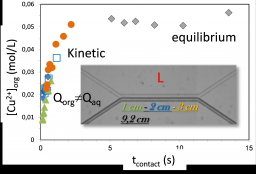
In the framework of an internal CEA program dedicated to the liquid-liquid extraction for nuclear waste treatment (or primary extraction of uranyl), we have fully characterized the structure of the organic phase with its influence on the phase stability[1],[2] and more recently on the extraction efficiency[3]. Extraction is related to solubilization properties[4] where surfactant and hydrotrope can be used in closed connection to enhance the transfer[5]. In such processes, the liquid-liquid interface plays a key role even though its specific influence is not fully understood. Recently, we took benefit of microfluidic tools to control the interface by making the interfacial reactions predominating over bulk ones. Dedicated home-made chips have shown the possibility to follow kinetic but also to control the shearing properties of the interface
[1] Chapter 7 for Ion Exchange and Solvent Extraction F. Testard, P. Bauduin, L. Berthon, Th. Zemb. Ed B. Moyer, CRC Press, chap 7, P381 (2009).
[2] Berthon L, Testard F, Martinet L, Zemb T, Madic C., C. R. Chimie (2010), 13 1326–1334
[3] Dourdain, S; Hofmeister, I; Pecheur, O; Dufreche, JF; Turgis, R; Leydier, A; Jestin, J; Testard, F; Pellet-Rostaing, S; Zemb, T Langmuir (2012), 28 11319-11328.
[4] Kunz, W., Testard, F., Zemb, Th., Langmuir, (2009), 25(1), 112-115.
[5] Bauduin, P., Zemb, Th., Testard, F., J. Phys. Chem. B (2008) 112, 12354-12360.
Supported lipid bilayers offer a unique configuration where a single bilayer, accessible to other molecules like, for example, proteins, peptides or DNA, is supported on a solid substrate. Beyond their interest for biosensor technology, the access they give to a flat immobilized membrane makes them highly relevant for fundamental studies in biophysics and membrane biology. In particular, they provide a unique way to finely characterize the interactions between membranes and their environment, which are not only crucial for membrane fusion and trafficking, endo- and exocytosis, but also fascinating from the physical point of view.
Membranes indeed exhibit extremely complex interactions with their environment, where molecular scale enthalpic and fluctuation related entropic contributions are inextricably involved. In particular, the effect of confinement has been now discussed for 40 years without finding a definitive answer. Helfrich (1973) first realized that, in addition to the “direct'' electrostatic, van der Waals and hydration forces, the long-range “effective'' steric interaction generated by the thermal fluctuations of confined flexible membranes is an essential contribution to the total free energy of interaction. Pure hard wall interaction (hard confinement) was first considered but is not a realistic description of real systems, and especially of living ones. Confinement by a “soft'' potential was treated either by using self-consistent methods leading to effective exponentially decaying potentials, or by estimating average values within a full statistical mechanics approach. Which functional form should be used to describe entropic repulsion in real experimental situations remains, however, an open question.
We have determined, by grazing incidence x-ray scattering, the interaction potential between two lipid bilayers, one adsorbed on a solid surface and the other floating close by[1]. We find that interactions in this highly hydrated model system are two orders of magnitude softer than in previously reported work on multilayer stacks. This is attributed to the weak electrostatic repulsion due to the small fraction of ionized lipids in supported bilayers with a lower number of defects. Our data are consistent with the Poisson-Boltzmann theory, in the regime where repulsion is dominated by the entropy of counter-ions. We also have a unique access to very weak entropic repulsion potentials which allowed us to discriminate between the various models proposed in the literature. We show that our data are best described using the soft potential of Podgornik and Parsegian (1992)[2]. We further demonstrate that the interaction potential between supported bilayers can be tuned at will by applying osmotic pressure, providing a way to manipulate these model membranes, thus considerably enlarging the range of biological or physical problems which can be addressed[3].
We are currently extending these measurements to the fluctuations and destabilization of membranes in an AC electric field.
[1] L. Malaquin, T. Charitat, and J. Daillant. Eur Phys J E Soft Matter, 31(3):285–301, 2010.
[2] A. Hemmerle, L. Malaquin, T. Charitat, S. Lecuyer, G. Fragneto, and J. Daillant. Proc. Nat. Ac. Sc. , 109(49):19938–19942, 2012.
[3] G. Fragneto, T. Charitat, and J. Daillant. EUROPEAN BIOPHYSICS JOURNAL WITH BIOPHYSICS LETTERS, 41(10):863–874, 2012.
Hybrid organic-inorganic nanoparticles with well-controlled morphology are currently of great research interest. Synthetic routes leading to robust aggregates made of nanoparticles of different chemical natures which are associated in a controlled manner, i.e. number of nanoparticles and geometrical arrangement, are especially investigated. Clusters of spheres could mimic the space-filling models of simple molecules and are called “colloidal molecules”. In the framework of the TOCOMO ANR project, the strategy is based on a seeded-growth emulsion polymerization process leading to biphasic particles, which are composed of spherical silica spheres (50-400 nm) surrounded by a varying number of polystyrene (PS) nodules.
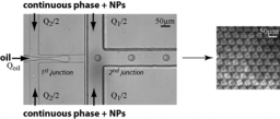
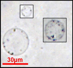
Emulsions of two immiscible fluids stabilized with solids particles, called Pickering emulsion, have been studied since the early twentieth century and the pioneering works of Ramsden[1] and Pickering[2] The irreversible adsorption of particles at the interface of the two phases gives original properties of these emulsions. Such emulsions are of primary interest in the food, cosmetics industry and oil recovery. Although it is now clear that the stabilization mechanism comes from the partial wettability of the nanoparticles by the two emulsion phases, the interface structures for such a stability mechanism is not well understood. There is a need of making a Pickering emulsion model system where the nanoparticles are well controlled as well as the emulsion size. The LIONS laboratory has developed a microfluidics platform (Fig.6) which allows preparing monodisperse tunable Pickering emulsion starting from monodisperse silica nanoparticles (NPs) of 7nm radius.
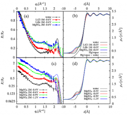
Within the ANR project ANR-06-BLAN-0276 "SISCI" (Spécificité Ionique dans les Systèmes Colloïdaux et Interfaciaux), we have explored the specific local ionic distributions in water in the vicinity of interfaces between aqueous electrolytes and external media.
Three different X-ray reflectivity techniques have been developed[1]. (i) The air-water interface has been investigated using grazing incidence x-ray fluorescence for different alkali-halide solutions[2]. The measured integrated amount of desorbed ions follows a Hofmeister series, in agreement with the surface tension behavior, and is interpreted in terms of ion-surface interaction potentials. (ii) The spatial ion distribution has been resolved at the sub-nanometer level using x-ray standing waves at the interface with a multilayer silica substrate. (iii) x-ray reflectivity analysis of polarized liquid mercury surface in contact with aqueous electrolyte has shown how the Hg-surface layering and the local ionic profiles depend on both applied potential and ion nature[3].
Carbonated mineralization as carried out by living organisms like seashells is a very intriguing process leading to crystalline materials that diffract like single crystals while adopting very sophisticated shapes. Nacre is a paradigmatic example of biomineralization, made of water-insoluble organic compartments filled with a CaCO3 mineral phase. We successively considered two model systems to mimic the influence of an organic template on mineralization: first, β-sheet peptide monolayers at the air-water interface and then giant unilamellar polymer vesicles.
Concerning the first model system, two different peptidic sequences, named LSFD and LDFD (respectively 12 and 11 amino-acid long), were designed and evaluated with respect to their ability to form β-sheet layers at the air-water interface[1] and to induce a specific CaCO3 crystallization by selecting the nucleated polymorph and/or its crystallographic orientation.[2] Although the two sequences exhibit a β-sheet conformation at the air-water interface and form 2D crystalline domains, they show unable to direct the mineralization: the selected crystalline polymorph is switched from calcite to vaterite when the interface is covered by any of the two peptidic films, but probably owing to a kinetic effect. Moreover, vaterite crystals appear as a 3D powder with no preferred orientation with respect to the organic “template”. Surprisingly, the Langmuir film made from the more acidic peptide (LDFD) remains stable upon mineralization whereas the LSFD peptide film gets destabilized, likely through an adsorption of the peptides on the growing crystallites.
Titanium and its alloys are widely used in aeronautical, marine and chemical industries for their good mechanical properties, high resistance to corrosion, especially in sea water or human body, and low density, about 60% of steel density. However, tribological properties of titanium surface are poor with highly unstable friction coefficients and adhesive wear mechanisms which lead mechanical parts to seize up. Surface treatments are required to improve surface properties, according to the envisioned application.
Nitriding and carburizing are the most commonly used thermochemical treatments, but they must be done in highly oxygen-free atmospheres to avoid titanium oxidation. This rules out the possibility of local treatments. Laser induced modifications offer an alternative tool for modifying the surface composition by insertion of light elements (oxygen, nitrogen, carbon) present in the reactive atmosphere. Modified layers must have a high content of nitrogen to be hard enough to stand up to surface friction, while oxygen, localized in less crystallized phases, is also needed to accommodate friction without failure. Oxynitride formation appears then as ideal to reach optimal resistance.
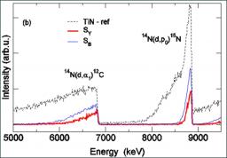
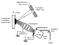
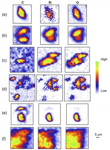
Extraterrestrial materials examined by mean of nuclear microprobe
H. Khodjaa,b, T. Smitha,b, C. Engrandc, G. Herzogd, C. Raepsaeta,b
Nuclear Instruments and Methods in Physics Research B 306 (2013) 245–248
- a CEA/IRAMIS/SIS2M/LEEL, F-91191 Gif-sur-Yvette, France
- b CNRS/UMR 3299/SIS2M/LEEL, F-91191 Gif-sur-Yvette, France
- c CSNSM, Bat 104, F-91405, Orsay Campus, France
- d Department of Chemistry and Chemical Biology, Rutgers University, 610 Taylor Road, Piscataway, NJ 08854-8087, USA
The LCCEF has been developing the amide chemistry for a long time due to the great chemical opportunities afforded by the NR2 ligands. In particular, the bis(trimethylsilyl)amide ligand (N* = N(SiMe3)2), ubiquitous in coordination chemistry, is able to stabilize metal centres in their lowest and highest oxidation states and can be substituted with proton acidic molecules, so that bis(trimethylsilyl)amide compounds are valuable starting materials for inorganic and organometallic syntheses. In the presence of a base the “MXN*” complexes are often transformed into metallacycles [M(κ2(C,N)−CH2SiMe2N{SiMe3})] following γ-CH deprotonation and HX elimination.
The parahydrogen induced polarization (PHIP) method is based on the non-Boltzmann distribution of nuclear spin states following the hydrogenation of a substrate by parahydrogen. Absorptive and/or emissive NMR signals can be greatly enhanced, up to several orders of magnitude as compared to thermally polarized molecules.
The research in this field is part of the expertise of LSDRM in its effort to increase NMR sensitivity.
Dihydrogen, initially made of 1/4 of parahydrogen and 3/4 of orthohydrogen, a proportion that does not induce any hyperpolarization, is cooled to -196°C in the presence of a ferric catalyst, thanks to an home-made apparatus. The resulting gas contains about one half of parahydrogen, and may be use to obtain hyperpolarized spectra.
A first example: the method named "signal amplification by reversible-exchange" (SABRE) is used here to increase the aromatic signals of N,N-dimethylnicotinamide. In SABRE, a catalyst reversibly binds dihydrogen and the aromatic molecule.
Nanostructuration is one of the most promising strategies in the quest for next-generation thermoelectric devices. This led us to study the thermoelectric conversion and its fluctuations using a scattering approach suitable for coherent quantum transport. Studying the thermopower of a chaotic quantum dot, we have found that a new family of universal distributions occurs when this is not the bulk but the edges of the dot spectrum which are probed at the Fermi energy. We have also studied disordered nanowires and calculated their thermopowers as well as their figures of merit and their maximal output powers to determine the optimal working conditions for the low temperature thermoelectric conversion.
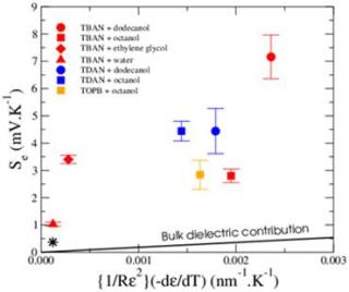
The shortage in fossil fuel resources has encouraged a huge effort towards the research of alternative sustainable energy. Within this framework, thermoelectricity offers the possibility to convert low-grade waste heat (including solar heating) into electric power. When a temperature gradient ΔT is applied to an isolated conducting rod, the electrons at the hot part acquire some kinetic energy and diffuse to the cold part, resulting in the creation of an electric field E=-ΔV=SΔT. The proportionality coefficient S is called “Seebeck coefficient”, from the name of the German physicist who discovered this effect. A tremendous effort is currently accomplished in the field of solid-state thermoelectric devices (more than 9,000 publications during the last decade). However, the research of new solid-state thermoelectric materials has reached its limits and most efforts are now devoted to the increase of the conversion efficiency through nanostructuration, which incurs a substantial manufacturing cost.
The recent discovery that spin angular momentum can be exchanged between the magnetization of an insulating ferromagnet and the conduction electrons of a normal metal layer has opened new perspectives [1]. In particular, it is now possible to incorporate materials such as YIG (Yttrium Iron Garnet, well known for its unsurpassed microwave properties) in innovative spintronic devices. Thanks to the recent progress in the growth of high quality, ultra-thin YIG films [2], electronic control of their relaxation obtained through pure spin currents generated by the spin Hall effect at the interface between a strong spin-orbit metal (e.g., Pt) and YIG might be possible.
Conversely, the spin current emitted by the magnetization precession can be used to probe the YIG dynamics [3,4]. This work is part of a new fruitful axis of research, "magnonics": the use spin waves (magnons) instead of electrons to transmit and process information. The idea is to nanopattern ultra-thin YIG films to take advantage of the specific properties of spin wave propagation in confined geometries and to propose new electronically controlled microwave devices. Magnetic Resonance Force Microscopy [5] is used to perform ferromagnetic resonance on magnetic nanostructures.
[1] Y. Kajiwara, et al., Transmission of electrical signals by spin-wave interconversion in a magnetic insulator. Nature 464, 262 (2010).
[2] O. d'Allivy Kelly, Hahn, et al., Inverse spin Hall effect in nanometer-thick Yttrium Iron Garnet/Pt system. Appl. Phys. Lett. 103,
082408 (2013).
[3] C. Hahn, et al., Comparative measurements of inverse spin Hall effects and magnetoresistance in YIG/Pt and YIG/Ta. Phys. Rev. B 87,
174417 (2013).
[4] C. Hahn, et al., Conduction of spin currents through insulating oxides. arXiv:1310.6000 (2013).
[5] B. Pigeau, C. Hahn, et. al., Measurement of the Dynamical Dipolar Coupling in a Pair of Magnetic Nanodisks Using a Ferromagnetic Resonance Force Microscope. Phys. Rev. Lett. 109, 247602 (2012)
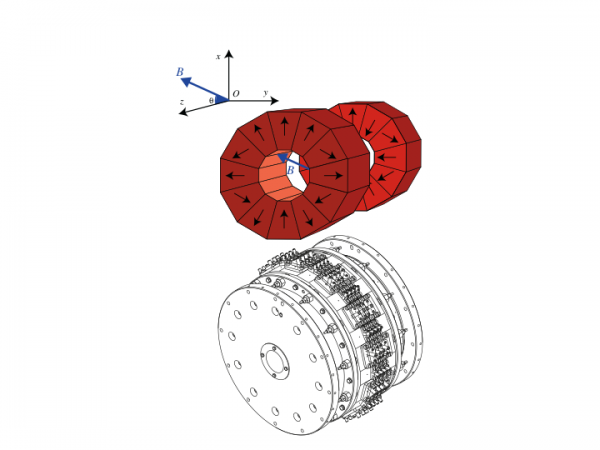
Les nano-oscillateurs à transfert de spin (dénommés ci-après STNO) sont des dispositifs de l'électronique de spin capables d'émettre une onde hyperfréquence monochromatique lorsqu'ils sont pompés par un courant polarisé en spin grâce au couple de transfert de spin (STT) [1,2] . Bien qu'ils offrentde nombreux avantages (agilité spectrale, intégrables sur un circuit onolithique, etc...), leur puissance d'émission est en général très faible.
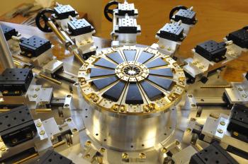
Une solution est de développer un réseau de STNO couplés, tous synchronisés entre eux. Dans ce cas, la puissance d'émission varierait en N² où N serait le nombre d'oscillateurs. Le but de cette étude expérimentale est d'étudier les différents types de couplages pouvant induire une cohérence de phase entre oscillateurs. On utilisera pour cela une méthode originale de détection de la résonance magnétique directement inspirée des techniques de microscopie en champ proche, la MRFM [3,4]. Le principe de détection consiste à utiliser un micro-levier mécanique avec une sphère magnétique accrochée à son extrémité pour faire de la spectroscopie locale par résonance magnétique. Le SPEC est l'un des deux laboratoires dans le monde qui maîtrise cette approche. Outre sa sensibilité (environ 100 spins) l'outil est aussi capable d'imager la dynamique dans des dispositifs avec une résolution spatiale nano-métrique.
Ce projet de recherche s'inscrit dans le cadre des projets européens Mosaic et ANR P2N Spinnova.
[1] D. C. Ralph & M. D. Stiles, Spin transfer torques, J. Magn. Magn. Mater. 320, 1190 (2008).
[2] A. Slavin & V. Tiberkevich, Nonlinear Auto-Oscillator Theory of Microwave Generation by Spin-Polarized Current. IEEE Trans. Magn. 45, 1875 -1918 (2009).
[3] O. Klein, et al., Ferromagnetic resonance force spectroscopy of individual submicron-size samples. Phys. Rev. B 78, 144410 (2008).
[4] A. Hamadeh, et al., Autonomous and forced dynamics in a spin-transfer nano-oscillator: Quantitative magnetic resonance force microscopy. Phys. Rev. B 85, 140408(R) (2012).
Magnetoresistance, giant or tunnel, is at the heart of several spintronic components, a new branch of electronics based on the spin of the electron with applications in information technology. Because of the never ending drive for miniaturization, spintronics is about to reach the ballistic regime of electronic transport for which spin dependent properties are still not completely understood. Further size reductions can even be achieved in constrictions of atomic sizes obtained by slowly stretching a nanostructure in a sensitively controlled manner. In these systems, static magnetotransport measurements result from the low-dimensional magnetism of a few relevant atoms. In parallel ferro-magnetic resonance (the magnetization precession induced by a radio-frequency magnetic field) has seen a renewed interest lately as it has been shown that magnetization dynamics interacts with spin currents. Its correlation with DC transport is being widely studied and it is now possible to electrically measure ferromagnetic resonance using the inverse spin Hall effect or the Anisotropic Magneto-Resistance (AMR).
The latter can be scaled down to atomic sizes thus giving the possibility to open the unknown field of the dynamical magnetic properties of a single atom in a low dimensional local geometry. We have very recently demonstrated that FMR can be detected in narrow constrictions using the AMR mixed with the RF current auto induced in the magnetic circuit, leading to a measurable DC voltage. After a strong experimental effort, an original setup has been built which allows us to electrically detect the ferromagnetic resonance at 77K in samples that can be broken in real time during the measurements. This unique tool will allow us to study the resonance properties of a single atom in a low-dimensional configuration. It may even allow us to demonstrate that some metals like platinum could become magnetic in these atomic contacts.
Post-doc financé par une bourse européenne "Marie Curie Intra-European Fellowship".
L’objectif principal de cette thèse consiste à développer une sonde à base de capteurs magnétorésistifs (Giant Magnetoresistance – GMR ou Tunnel Magnetoresistance - TMR) afin de mesurer le champ magnétique induit par les courants circulant le long des neurones. L’électrophysiologie classique permet actuellement d’enregistrer les potentiels électriques locaux des cellules nerveuses mais les électrodes utilisées n’apportent pas d’informations sur la signature magnétique de ces cellules. La sonde (appelée "magnétrode" en référence aux électrodes d’électrophysiologie) doit permettre d’obtenir la réponse électromagnétique des neurones, en utilisant la très bonne sensibilité et la possibilité de miniaturisation des capteurs magnétiques très sensibles (GMR ou TMR).
Dans un premier temps, la fabrication, la miniaturisation et la caractérisation des magnétrodes seront effectuées puis des tests in-vitro et in-vivo seront réalisés en collaboration avec respectivement l’équipe d’Alain Destexhe et Thierry Bal de l’Unité de Neurosciences, Information et Complexité (UNIC) du CNRS à Gif-sur-Yvette et celle du Professeur Pascal Fries de l’Ernst Strüngmann Institute (ESI) for Neuroscience in Cooperation with Max Planck Society à Francfort.
Magnetization dynamics in individual vortex-state nanodots has been investigated using the spectroscopic technique of magnetic resonance force microscopy (MRFM) developed in the LNO. It has led to the discovery of future potential magnetic memories based on the resonant switching of the vortex core
A magnetic vortex corresponds to a curling in-plane magnetization distribution leaving a small core region (a few nanometers wide, the size of the exchange length) at the center of the dot, where the magnetization is pointing out-of-plane, either up (polarity p=+1) or down (p=-1). Its lowest energy mode corresponds to a gyrotropic motion of the vortex core about its equilibrium position. This mode has recently attracted practical interest for future magnetic memories and microwave devices by allowing the engineering of the spin-wave excitation spectrum of isolated nanodots and large array of dipolarly coupled nanodots (magnonic crystals).
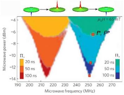
We have first demonstrated that the frequency degeneracy corresponding to the gyrotropic modes with opposite polarities in zero field can be lifted by applying a magnetic field perpendicular to the disk plane [1] This Zeeman-like splitting can be used for a simple reading of the polarity state in an individual nanodisk. In order to discriminate the resonant frequencies f- and f+ associated respectively to the core polarities p=-1 and p=+1, it is necessary to choose the static magnetic field in such a way that the field-induced gyrotropic frequency splitting exceeds the linewidth of the gyrotropic mode. In our experiment, a bias field as small as µ0H=13 mT is sufficient to fulfill this condition.
We have proposed to take advantage of this frequency discrimination in order to reverse deterministically the vortex core polarity, as shown in references [2]. Starting with the vortex core polarity in, say, the p=+1 state, a single microwave field pulse whose carrier frequency is tuned at f+ and with sufficient amplitude will resonantly excite the gyrotropic motion of the core until it reaches a critical threshold for reversal (see Figure 1). Once it has been reversed, the final state p=-1 is out-of-resonance with the writing pulse so that it cannot be switched back to p=+1. Similarly, it is possible to write the state p=+1 starting from the p=-1 state using the appropriate pulse. This writing process has been shown to be very robust, as no mistake could be recorded out of several hundred attempts with our experimental parameters.
The multiband nature of the iron-based superconductors is a key factor to understanding their physical properties. We have performed a complete study of the charge transport in Ba(Fe1-xCox)2As2 single crystals, in connection with a determination of their electronic structures by photoemission experiments.
In the new Fe-based pnictide superconductors discovered in 2008, the appearance of high-Tc superconductivity in close proximity to the antiferromagnetic phase has been taken as the signature of unconventional superconductivity. It seems now well established that magnetism and superconductivity are directly connected to the peculiar features of the electronic structures of these compounds, which are characterized by small hole and electron pockets. The transport properties of the prototypical Ba(Fe1-xCox)2As2 have been studied throughout the electron doped phase diagram as fine tuning of Co content can be achieved in sizeable single crystals.
L'expérience dite de « von Karman » est une expérience consistant à produire un écoulement fluide dans un cylindre à l'aide de turbines munies (ou non) de pales. Ce sujet de recherche a connu un intérêt important dès les années 1990: l'ajout de pales aux turbines a permis d'atteindre des nombres de Reynolds et des taux de turbulence élevés ce qui a permis de vérifier directement plusieurs hypothèses fondamentales en turbulence (intermittence, hypothèses de Taylor, mesures de dissipation).
Cette expérience est utilisée au sein du laboratoire, qui possède une expérience importante de ce type d'écoulements, afin d'observer les diverses brisures de symétrie qui sont induites à la transition vers la turbulence et après cette transition. Les symétries du montage (axisymétrie, symétrie par inversion) permettent de traiter théoriquement le problème en effectuant une analogie avec les résultats de la turbulence à deux dimensions.
Cet écoulement est aussi étudié expérimentalement: deux techniques de visualisation (Vélocimétrie Laser Doppler et Vélocimétrie par Imagerie de Particules) révèlent des propriétés physiques complémentaires sur la turbulence produite par la rotation des turbines.
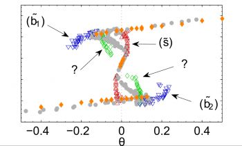
Cette expérience a mis à jour un grand nombre de résultats théoriques et expérimentaux: le système présente une Beltramisation (alignement vorticité / vitesse) à haut nombre de Reynolds, un hystérésis qui dépend des propriétés du forçage à haut Reynolds, et une éventuelle multi-stabilité rappelant les systèmes à basse dimension pour des conditions de forçage bien précises.
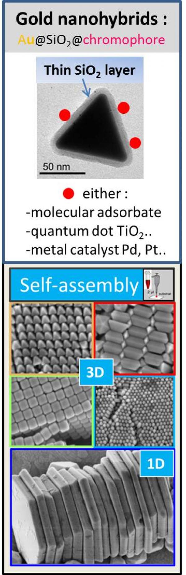
Responsable : Sylvie MARGUET
Participants: Aurélie Habert, Jérôme Caron (Master-2), Mohammad Khaywah (Post-doc)
IRAMIS-NIMBE-LEDNA (Laboratoire Edifices Nanométriques)
Résumé: notre objectif est de tirer parti de l'interaction lumière-matière dans des nano-hybrides, constitués de nanoparticules d'or (AuNPs) colloïdales, dont la morphologie est optimisée, pour générer de la lumière, de la chaleur ou des porteurs de charge, selon l'application visée*.
* ce thème est en lien avec ceux développés au sein de deux groupements de recherche (GdR) du CNRS en France. Les GdR Or-nano et GdR PMSE (Plasmonique Moléculaire et Spectroscopies Exaltées).
… pour la PLASMONIQUE: depuis 2008 :
Collaborations : CEA-SPEC; L2n-Troyes; ISMO-Orsay; IS2M-Mulhouse; ILM-Lyon, ..
La plasmonique est une discipline à l'interface entre la physique, la chimie et la biologie. L’excitation de la résonance plasmon d’une nanoparticule (NP) métallique permet de générer des champs électromagnétiques très intenses, confinés à la surface de la NP, qui peuvent être utilisés pour exalter ou initier une réaction photochimique dans un « chromophore » placé à proximité. Le confinement du champ électromagnétique autour d’une NP colloïdale, monocristalline et non rugueuse, est supérieur à celui autour du même objet fabriqué par nanolithographie. La relaxation du plasmon, qui accompagne cet effet d’amplification, se fait selon des voies de relaxation ultra-rapides, en compétition les unes avec les autres. Leur importance relative dépend, de la morphologie de la NP, de son environnement "proche" et du mode d'irradiation. Ainsi, les NPs d’or peuvent se comporter comme des nanosources de lumière, de chaleur ou de porteurs de charges (électron, trou), selon la voie de désactivation qui prédomine. Pour ce qui est de la morphologie, c'est la taille, la présence de pointes et le rapport d'aspect (surface/volume) de la NP qui comptent. L’environnement désigne à la fois les «chromophores» adsorbés à la surface de la NP et notamment leurs niveaux d'énergie (HOMO-LUMO pour une molécule ou VB-CB pour un semi-conducteur) ainsi que la présence éventuelle d’une couche à l’interface (résidus de surfactant, couche isolante..). Enfin, le mode d'irradiation, continu ou pulsé à différentes échelles de temps de la micro- à la femto-seconde, est aussi un paramètre clé pour l’observation de ces trois types de nanosources.
Nous synthétisons des NPs d’or (Au-NP) en contrôlant minutieusement leur morphologie (taille, forme) et leur structure cristalline. Certaines de ces AuNPs ne sont produites que dans quelques laboratoires à travers le monde. Les microplaquettes (triangulaires, hexagonales ou en forme de disque), atomiquement planes, sont prometteuses pour la nanofabrication FIB de motifs monocristallins, non accessibles autrement. Un grand nombre de protocoles sont en cours d'élaboration pour des finalités diverses : i) remplacement du surfactant initial par d'autres molécules plus appropriées ; ii) dispersion homogène des NPs sur divers supports ; iii) auto-assemblage en une (1D), deux (2D), ou trois dimensions (3D) pour fabriquer des « points chauds » ; iv) enrobage par une couche de silice d'épaisseur variable. Des applications liées à l’environnement et à l’énergie sont envisagées, à plus long terme, via la synthèse de nanoparticules multimétalliques du type Au@X (avec X= Pd, Pt, Au, Ag or TiO2) qui combine un composant plasmonique (Au) et un catalyseur pour la catalyse plasmonique et la nanophotochimie induite par plasmon.
… pour la MEDECINE : depuis 2018 :
Collaborations: LPQM-CentraleSupelec, LAC-Orsay, CEA-SPEC, PPSM-ENS, ..
Nous produisons des nanoparticules cœur-coquille Au@silice qui ont un fort potentiel comme agent de contraste pour de nombreuses techniques d’imagerie médicale (photoacoustique, diffusion champ sombre, luminescence multi-photonique, Ultrasons à haute fréquence, contraste de phase quantitatif, tomographie par ordinateur…). En ce sens, le projet ANR SINAPSE, récemment accepté vise à développer une nouvelle classe de NP pour l'imagerie neuronale (coord F. Marquier). Les NPs d’or sont d’excellents capteurs (e.g test de grossesse) du type LSPR, SERS, Fluorescence, Pression ... Dans le domaine de la photothérapie, les NPs d'or présentant un rapport d'aspect (longueur/épaisseur) élevé, tels que les fils et les plaquettes, sont très intéressantes car elles peuvent être excitées par des photons infrarouges, dans les premières fenêtres (I,II,III,IV) de transparence des tissus biologiques. La génération de chaleur induite par plasmon (PTT ; photothermal therapy) et plus récemment la génération de R.O.S. (reactive oxygen species) à partir de NPs d’or seules (c.a.d. sans photosensiblisateur) est une approche originale pour traiter les tumeurs, en particulier en absence d'oxygène, lorsque la PDT (photodynamic therapy) conventionnelle, basée sur l’utilisation de molécules type porphyrine à l'état triplet excité triplet, ne peut pas fonctionner. Le projet HEPPROS (plan cancer) a pour but d’étudier la génération de chaleur et de R.O.S. telles que 1O2, OH., O2.-, H2O2 pour diverses morphologies de NPs d’or et ainsi mieux comprendre les mécanismes transitoires complexes impliqués à l'interface AuNP/adsorbat moléculaire (coord B. Palpant).
Heterogeneous samples are usually studied using solid-state NMR methods and while the sample is spinning at the magic angle. This is problematic for many cases, where the sample cannot be spun, as in MRI. The alternative methodology of spinning the magnetic field around a static sample has been proposed since the 60's, but not yet successfully applied. Our goal is to develop the methodology and the instrumentation in order to perform magic angle field spinning and answer questions related to the monitoring of metabolism in living matter as well as the structure and function of complex systems like batteries, porous media and fuel cells.
Alexandre Hervé, Pierre Thuéry, Michel Ephritikhine, Jean-Claude Berthet
L’ion cyanure CN− est certainement l'un des ligands les plus utilisés en chimie de coordination des métaux de transition d. De nombreuses études ont montré l’intérêt des complexes cyanures dans des domaines très variés allant de la biologie aux nanomatériaux. En plus d’une riche chimie de coordination liée aux divers modes de coordination du group CN, les complexes cyanures présentent bien souvent de remarquables propriétés magnétiques, photoluminescentes et/ou de conductivité. Au contraire de celle des métaux d, la chimie des cyanures des métaux f (lanthanides et actinides) n’a été que très peu considérée.
Le laboratoire LCCEf s’intéresse depuis quelques années au développement de cette thématique à partir de précurseurs organométalliques ou comportant des ligands azotés, et a mis en évidence le potentiel du ligand CN−,
- pour stabiliser les nombreux degrés d’oxydation de l’uranium1-4
- et former des espèces inattendues (métallocènes linéaires,2,3 complexe cyclopentadiènylique de l’uranyle4) bouleversant ainsi les mentalités et ouvrant de nouvelles possibilités notamment en chimie organométallique.
Les études actuelles concernent la formation de briques moléculaires mono ou polycyanures, intéressantes pour de nouveaux développements en chimie de coordination, mais surtout susceptibles de conduire à la formation de clusters et d’agrégats homo ou polymétalliques originaux dont les propriétés photochimiques (magnétisme et luminescence) seront étudiées. Ces études expérimentales couplées à des calculs théoriques favoriseront également une compréhension plus fine de la liaison Actinide-ligand nécessaire sur les plans fondamental et appliqué (Nucléaire).
The LCCEF has been developing the amide chemistry for a long time due to the great chemical opportunities afforded by the NR2 ligands. In particular, the bis(trimethylsilyl)amide ligand (N* = N(SiMe3)2), ubiquitous in coordination chemistry, is able to stabilize metal centres in their lowest and highest oxidation states and can be substituted with proton acidic molecules, so that bis(trimethylsilyl)amide compounds are valuable starting materials for inorganic and organometallic syntheses. In the presence of a base the “MXN*” complexes are often transformed into metallacycles [M(κ2(C,N)−CH2SiMe2N{SiMe3})] following γ-CH deprotonation and HX elimination.

Formation of organo-compounds involving [Mf]=C double bond and description of the nature of this formally double bond is a subject of considerable interest for some years. The LCCEf, in collaboration with the Ecole Polytechnique, focused on the interaction of the novel dianionic carbene SCS2- with uranium ions.
Conditions were found which permit the use of UCl4 instead of the less convenient borohydride starting material U(BH4)4 to prepare the mono-, bis- and tris- uranium(IV) carbene complexes with the dianionic ligand [C(Ph2PS)2]2− (SCS2−) (Fig . 1).
Changing the geometry of a metallocene complex from linear to bent or vice versa is of importance for the control of physicochemical properties and/or reactivity and for the evaluation of the covalency and steric interactions in the metal-cyclopentadienyl bonding.
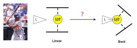
The parahydrogen induced polarization (PHIP) method is based on the non-Boltzmann distribution of nuclear spin states following the hydrogenation of a substrate by parahydrogen. Absorptive and/or emissive NMR signals can be greatly enhanced, up to several orders of magnitude as compared to thermally polarized molecules.
The research in this field is part of the expertise of LSDRM in its effort to increase NMR sensitivity.
Dihydrogen, initially made of 1/4 of parahydrogen and 3/4 of orthohydrogen, a proportion that does not induce any hyperpolarization, is cooled to -196°C in the presence of a ferric catalyst, thanks to an home-made apparatus. The resulting gas contains about one half of parahydrogen, and may be use to obtain hyperpolarized spectra.
A first example: the method named "signal amplification by reversible-exchange" (SABRE) is used here to increase the aromatic signals of N,N-dimethylnicotinamide. In SABRE, a catalyst reversibly binds dihydrogen and the aromatic molecule.
