

1. HR-MAS of micro-scale specimens
1H HRMAS (High-Resolution Magic-Angle Spinning) NMR spectroscopy has found success in the study of metabolome in heterogeneous biospecimens, such as cells, tissues and orgamis, owing to its sample non-destructive nature and simplicity data aquisition. However, NMR in general is an insensitive spectroscopic technique relying on large sample quantity, typically 10-20 mg per spectral data; and it has limited the metabolic invertigations in biology and in medicine. For this reason, there is presently a need to develop new NMR methodologies that are capable for analyzing small-scale specimens. One approach is the use of a micro-size detection under MAS condition, but unlike the standard uMAS commerical probe, it must offers high detection sensitivity without compromising the spectral resolution (i.e., <0.002ppm). Here, we are developing a HR-MAS methodology capable of offering excellent spectral quality in sensivity and resolutoin for metabolic applications.
References:
'Evaluation of a high-resolution micro-size magic angle spinning (HRuMAS) probe for NMR-based metabolic studies of nanometer samples'
N. T. Duong, Y. Endo, T. Nemoto, H. Kato, A-K Bouzier-Sore, Y. Nishiyama, A. Wong, Anal. Methods 8 (2016) 6815-6820.
'High-resolution NMR-based metabolic detection of microgram biopsies using a 1 mm HRuMAS probe'
Y. Nishiyama, Y. Endo, T. Nemoto, A-K Bouzier-Sore, A. Wong. Analyst 140 (2015) 8097-8100.
Financial Supports:
- ANR-PRC (2016-2019) on HRuMAS.
- ANR-JCJC (2012-2015) : Développement et exploration de nouveaux micro-détecteurs RMN tournants pour des analyses métabolomiques de biopsies de petite masse – HRMACS
2. Spatial Localized MAS NMR Spectroscopy
The standard HR-MAS NMR of biospecimens is a very promising technique for metabolic analysis of unprocessed and heterogeneous specimens; however, the obtained metabolic data represent only the bulk sample, with the lost of spatial localized information. The recent introduced localized-MAS NMR technique – by integrating a MAS probe with a standard micro-imaging gradient system – enables to profile the metabolite contents in different body region of a small organism, without independently analyzing the different body components. The benefit of the experimental setup is that the gradient coils (along x, y, z) are not implemented onto the MAS stator and are separated from the detection rf coil, minimizing any possible disturbances on the rf coil; also, it eliminates any torque acts upon the MAS stator during the gradient pulses, and thus mechanical stability is not compromised. Moreover, the effective gradient strength along with magic-angle GMAS is √3Gx,y,z, in which it is generated by simultaneously superposing three equal components of Gx = Gy = Gz when the MAS rotor axis is oriented at a 45° of the X-Y laboratory frame (Euler angle α = 45°).
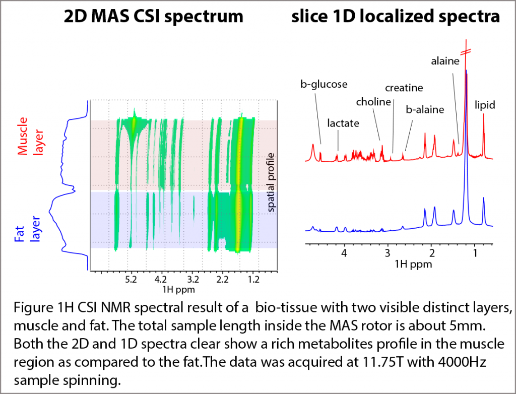
The figure shows the capability of CSI (Chemical Shift Imaging) spectra for separating the two different regions of a bio-tissue – fat and muscle layers. The 1D sliced spectra reveal a relatively rich metabolite content (between 3-4 ppm) inside the muscle layer as compared to the fat region.
Presently, the localized-MAS spectroscopy is at a primitive stage of the development. We are exploring explore the experiment, in hope to enhance spectroscopic potential towards metabolomic-based studies.
Reference:
'Metabolite localization in living drosophila using high resolution magic angle spinning NMR'
Sarou-Kanian V, Joudiou N, Louat F, et al. Sci Reports 5 (2015) 9872.