Photoionization time delays measured with coincidence spectroscopy
The availibity of attosecond pulses in the XUV domain has triggered the study of photoionization timings (see e.g. the review of M. Dahlström et al.) . Depending on the potential it rides on the way out, an electron may appear in the continuum earlier or later. This kind of delays was first measured in atoms by the groups of MPQ in Garching and Lund University) far from resonances, while we investigated the role of resonances in nitrogen in 2009-2011.
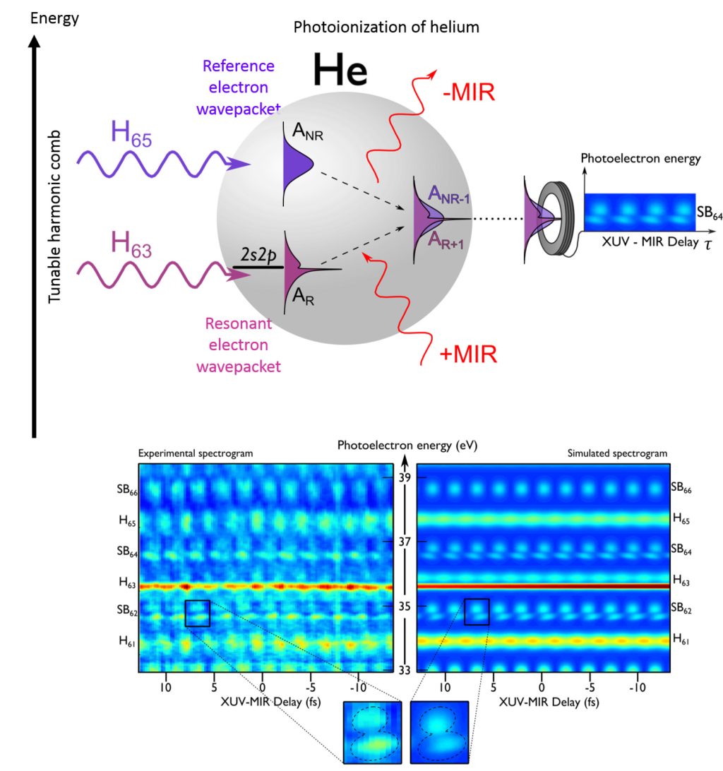
Here we implemented the RABBIT technique in He target atoms, and focused our investigations on the spectral region of its Fano resonance close to 60.15 eV. We could tune the wavelength and spectral width of our XUV harmonic comb thanks to the insertion of an OPA stage between the Ti:Sapphire laser and the HHG cell. Using a time of flight spectrometer with a high spectral resolution and generalizing the RABBIT approach to intra-sideband analysis, we could reconstruct the build up of this resonance profile against time, and show how it converges to the 17 fs lifetime long-known Fano profile.
This research was performed on PLFA laser in Saclay for its experimental part, in a collaboration between the Campus group in Madrid, and LCPMR (Pierre and Marie Curie University in Paris) . See also the press release on the CEA website Gruson et al. and that of Madrid University Gruson et al. in spanish.
Photoionization of rare gases by a train of attosecond pulses in presence of a dressing IR field: coincidence measurements
Attosecond light sources, which are emitting in the XUV spectral range, are naturally adapted to studies of photoionization of any species. Due to low XUV fluxes, studies have been mostly limited so far to IR+XUV pump/probe schemes, where it is the value of the vector potential associated to the IR-field that is providing the probe parameter. This process of photoionization by a combined action of an XUV and IR pulses synchronized within a fraction of their period forms the basis of temporal characterization methods: FROG-CRAB (or streaking) which uses an intense IR field is dedicated to isolated or to a few attosecond pulses, while RABBIT, which uses a low IR intensity, is dedicated to pulse trains.
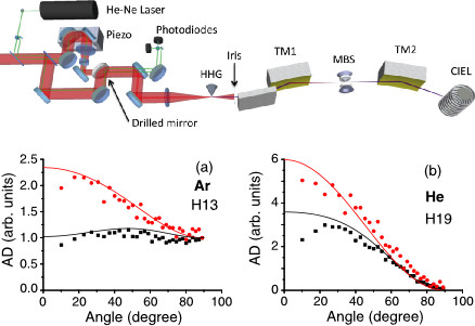
Here, we carefully investigated the whole regime from low dressing to large dressing values, using a coincidence imaging detector. Traces of photoionization of argon and helium gases angularly resolved for a series of sidebands and harmonics were recorded in coincidence, using a cold target recoil ion momentum spectroscopy (COLTRIMS) apparatus. We observed that whereas angularly resolving the sidebands does not help to investigate the process, the shape of the « dressed » harmonics was very sensitive to the dressing intensity and delay. This was reproduced by theoretical calculations.
This research was performed on PLFA laser in Saclay for its experimental part, in a collaboration between ISMO (Paris Sud University), and LCPMR (Pierre and Marie Curie University in Paris) .
Accessing photoionization timings
Related publications :
When performing an XUV/IR cross correlation in a target gas with an attosecond pulse train made of odd harmonics of the IR frequency, it is predicted that the photoionized electrons will spread over main lines and sidebands. Both will oscillate with the delay between the XUV and the IR beam, with half an IR period. This process, which is at the basis of the RABBIT technique (see Equipments & methods page) has long been used with a known target species to access the properties of the XUV radiation, in particular its spectral phase.
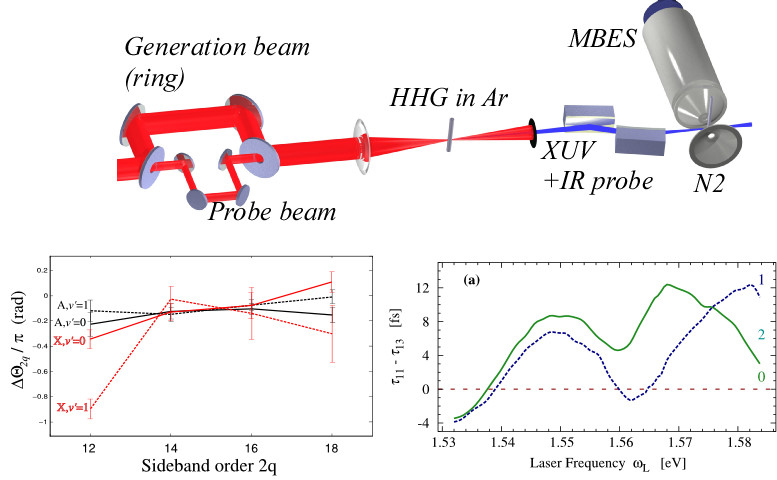
In a series of experiments and simulations, we showed that the exact same protocol may be used to probe the photoionization phase of an unknown species. This was illustrated on molecular nitrogen which showed an unexpected phase behavior when an harmonics was resonant with a Hopfield Rydberg state. Scanning the wavelength of the driving IR field, it was shown that the arrival time of the electronic wave packets on the detector could be accessed. This is very much related to the study of photoionization group delays as studied in Lund ((Atomic Physics Department in Lund – Sweden) and Garching (Attosecond Physics Laboratory at the Max Planck Institute of Quantum Optics in Garching – Germany) ) but in the molecular case.
This research was performed on PLFA laser in Saclay for its experimental part, in a collaboration LCPMR (Pierre and Marie Curie University in Paris).
Angularly resolved electronic dynamics (work carried out in Lund)
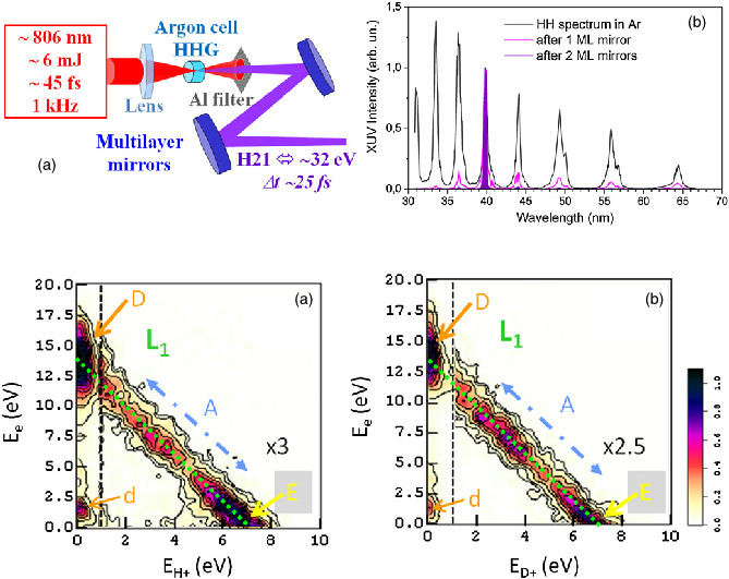
We used HHG as a secondary XUV and femtosecond source to study photoionization of H2 and D2 molecules close to the resonance of doubly excited states Q1 and Q2. We selected a single harmonic with a series of tailor made multilayer XUV mirrors provided by our colleagues from Institut d’Optique Graduate School, centered about 32 eV (38nm). The pulse duration was estimated to be 20 fs with a driving laser pulse provided by the PLFA server in Saclay, at 1 kHz repetition rate . The dissociative photoionization of H2 and D2 was studied in a cold target recoil ion momentum spectroscopy (COLTRIMS) apparatus, finally giving the molecular frame photoelectron angular distribution of the atomic fragments. It was shown that they compare very well to previous data obtained on a synchrotron high repetition rate source. It opens the door to time resolved experiments using MFPAD at ultimate time scales.
This research was performed on PLFA laser in Saclay for its experimental part, in collaboration with our colleagues from ISMO (Paris Sud University).
Molecular frame photoelectron emission for dissociative photoionization
Related publications :
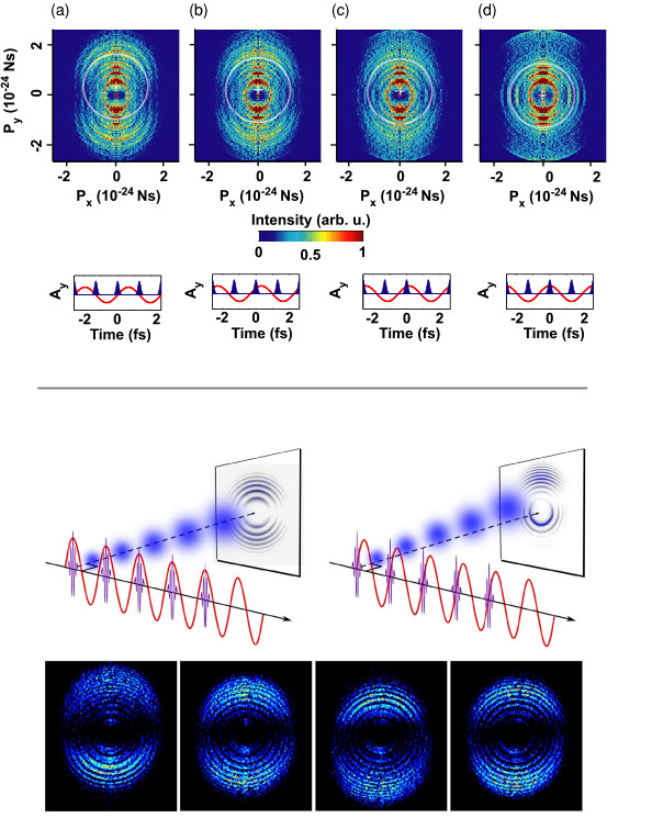
The RABBIT process, as described in the Equipments & methods page, may be performed with any electronic spectrometer, the only limitation being the possible angular integration of the detector. In standard experiments, to benefit from an intense signal, the signal is fully integrated over 2π in a magnetic bottle electron spectrometer. At the price of a great experimental effort due to the relatively low repetition rate of our lasers, it was performed in a full 3D imaging spectrometer ( COLTRIMS, see above). As an intermediate step, we may image in 2D systems that possess an axial symmetry using a velocity map imaging spectrometer (VMIS). It offers a fairly high counting rate making XUV-IR cross-correlation accessible in a reasonable time.
We performed this kind of experiments with two different attosecond pulse trains using a fairly intense dressing IR. In this regime, the photoelectron created by the attosecond pulse train is driven by the dressing laser in an oscillatory motion away from the atom and its final velocity, which determines its impact position on the detector, is solely determine by the time at which it has been ionized. In practice, the vector potential (A) associated to the IR field is the key parameter. Using two successive XUV attosecond pulses synchronized with a maximum or a minimum of A, we could shift in opposite directions two wavepackets. Their interference on the detector revealed the phase of their electronic wavefunctions.
In a second series of experiment, we synthesized attosecond pulse trains which pulses could all be synchronized with the same vector potential. In that case, all electronic wavepackets end up at the same location on the detector, which depends on the relative timing of the XUV vs. the IR. Scanning this delay, an « electron movie » is obtained. Information may be retrieved on the scattering of the electron on the ionic core on its oscillatory way out.
This research was performed in Atomic Physics Department in Lund – Sweden.


