Contact CEA : Hamed Merdji
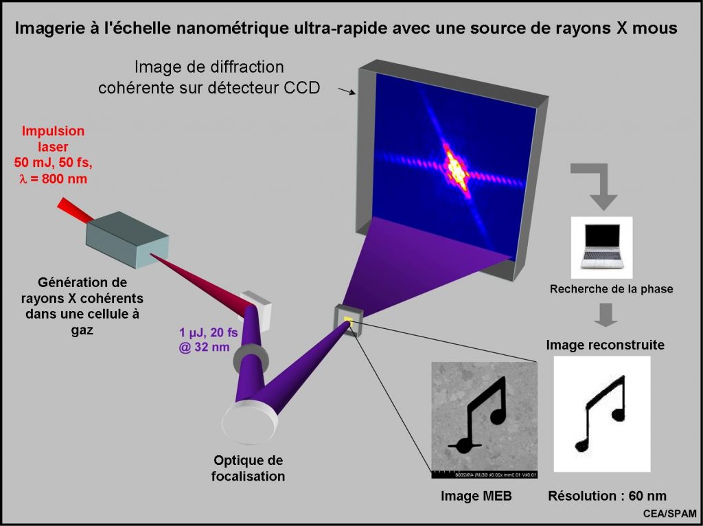
In photography, the scattered light from an illuminated object is recorded with a detector and one get an image of it. If the image is formed with an objective, the optics imposes many limitations (resolution, aberrations …). To achieve the ultimate resolution, spatially (function of the wavelength of the radiation used) and temporally (function of the “flash” duration), one possible technique (without any optics) is the coherent diffraction. Using a coherent beam like a laser to illuminate the object, a signal modulation due to interference is present and allows digitally reconstructing the exact image of the object with an unprecedented precision. To achieve nanometer or even atomic resolution, we therefore enlighten with a beam of coherent X-rays (radiation laser wavelength nanometer) and record the image. The usually low average illumination requires long accumulations over several laser shots. Recent advances have yielded images with a single shot femtosecond (10-15 s) from a laser laboratory, opening the way for time resolved studies.
For regular arrangements of elementary objects, the Bragg diffraction of X-rays is a powerful technique for characterization of matter at the atomic scale. It is the primary tool for crystallography. The information contained in the Bragg diffraction is rich: if a is the characteristic size of the elementary object, the Bragg peaks are spaced by 1/a in reciprocal space. However, some information is lost: indeed, the maximum frequency at which the diffraction pattern can be sampled is less than the Nyquist frequency (2a). In particular, if the elementary object has an amplitude and a phase, the phase is lost in the Bragg diffraction.

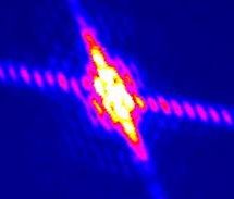
While the Bragg diffraction is limited to periodic arrangements of objects, coherent diffraction imaging allows to record the amplitude and phase for extended and isolated non-periodic objects. Indeed, the continuous diffraction pattern (Fig. 1) allows an “oversampling” of the pattern at a frequency above the Nyquist criterion, allowing the complete reconstruction of the amplitude and phase profiles of the object. The coherent diffraction in the extreme UV (XUV) and the X requires a highly coherent beam(transverse coherence) that has been recently produced. Early studies of “Coherent Diffraction Imaging” (CDI) have used the synchrotron radiation 3G (Miao Nature 1999). The applications were then extended to biological objects and nano-particles. Recently, Chapman et al. (Nature Phys. 2006) have imaged single nanoscale objects, using ultra-short single pulses (length = 30 nm, duration ~ 20 fs), issued by the free electron laser FLASH (TGI). This demonstration of imaging “in single shot” opens important perspectives in imaging isolated non-periodic systems (eg non-crystallisable), such as isolated proteins, cells, viruses or nano-particles.

Within SPAM, the attophysics team of Hamed Merdji offered to meet the challenge of CDI on an ultra-fast harmonic source [1], a priori less brilliant than FLASH, but much more compact (about 10 m against 350 m). The CDI beam line installed on the LUCA laser is illustrated in Fig. 1. The harmonic radiation generated in a gas is selected spectrally (pulse length = 32 nm, energy ~ 1 μJ, duration 20 fs) and focused (~ 4 microns beam diameter) on the object by an off-axis parabola. The object amplitude (Fig. 2a) is etched by a focused ion beam on a 150 nm thick membrane (collab. Lab. Photonics and Nanostructures). Fig. 2b shows the diffraction pattern obtained in only 40 shots, and the reconstruction of the object. The effective resolution (~ 62 nm) is consistent with the expected value (R=l/NA ~ 30 nm, NA=D/L numerical aperture). Note that the only published CDI studies using the harmonic source (Sandberg PRL 2007) require about one million shots! The team in Saclay has continued its efforts and obtained a CDI with one single shot (Fig. 2c), matching the performance of FLASH on a laser table.
Recent developments have allowed us to optimize the single shot CDI. This allows us to investigate now time-dependent systems, such as the dynamics of magnetization at the nanometer and femtosecond scales (collab. J. Luning, LCPMR, ANR 2009 Femto-X-Sp).

References:
[1] Single-shot diffractive imaging with a table-top femtosecond soft X-ray laser-harmonics source
A. Ravasio, D. Gauthier, F. Maia, M. Billon, J-P. Caumes, D. Garzella, M. Géléoc, O. Gobert, J-F. Hergott, A-M. Pena, H. Perez, B. Carré, E. Bourhis, J. Gierak, A. Madouri, D. Mailly, B. Schiedt, M. Fajardo, J. Gautier, P. Zeitoun, P. H. Bucksbaum, J. Hajdu and H. Merdji,
Phys. Rev. Lett. 103(2) (2009) 028104.
Margaret M. Murnane & Jianwei Miao
News and Views, Nature 460, (2009) 1088.


