


Pages scientifiques 2008
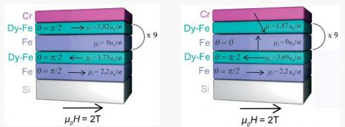
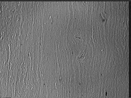
The magnetization of [Fe 3nm/Dy 2nm] multilayers has been studied. The samples were thermally evaporated under ultra-high vacuum at different substrate temperatures varying from 320 K to 870 K. In order to get the magnetization depth profile of these Transition Metal/Rare Earth (TM/RE) multilayers, an investigation of the structural, chemical, and magnetic properties was carried out. The samples were studied by High Resolution Transmission Electron Microscopy (HRTEM), Three-Dimensional Atom Probe (3DAP) and Polarized Neutron Reflectivity (PNR). The multilayers have been found to be rather homogeneous, except for the first two bilayers deposited on the substrate : the mainly crystalline structure of the first Fe layers leads to an enhancement of the ordering temperature of amorphous Dy. Moreover, at low temperature, a negative exchange coupling between Fe and Dy layers has been evidenced. Magnetization profiles have also been calculated by Monte Carlo simulations to support the PNR fits.
Quantum Shot Noise: the silence of the electrons
- Absence of shot noise for perfectly transmitted electrons:
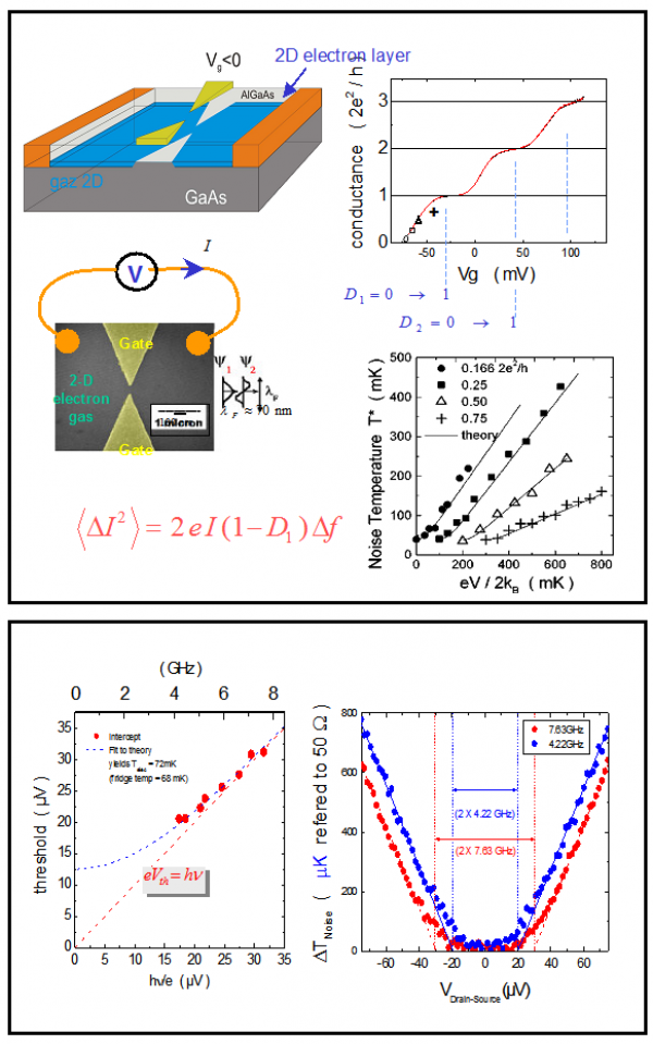
Fermi statistics (or Pauli exclusion) is responsible for the regular (noiseless) emission of electrons by a contact biased at potential V. Electrons are emitted at frequency eV/h in each mode able to propagate through the conductor. For perfectly transmitted electronic mode no current noise is observed while if the mode is partially transmitted, transmission/reflection of electrons occurs leading to current fluctuations. This quantum shot noise arise both from electron granularity and quantum uncertainty. The Nanoelectronics Group has been pionneer in developping noise correlation techniques in order to observe the full quantum shot noise suppression in a Quantum Point contact and provide quantitative comparison with quantum scattering models. 'Experimental Test of the Quantum Shot Noise Reduction Theory', A. Kumar, L. Saminadayar, D. C. Glattli, Y. Jin, and B. Etienne, Phys. Rev. Lett. 76, 2778-2781 (1996)
- Photo-assisted quantum shot noise:
Can one measure electron shot noise while no current flows through a conductor? We showed that the role of the bias voltage can be replaced by the photon energy quantum hn when the contact is irradiated by a microwave of amplitude Vac. An electron emitted by the contact may absorb a photon with probablity ~(eVac/2hn)2 and an electron-hole pair is created. Electrons and holes are separately transmitted/reflected through the conductor therfore generating current fluctuations or shot noise even though no dc current can be generated by the neutral e-h pairs. Using a Quantum Point Contact we provided direct quantitative measurements of photo-assisted e-h shot noise and its complete suppression when electronic mode are perfectly transmitted. Also, upon applying a dc bias voltage V, the competition between transport and photo-assisted noise leads to an observable singularity in the noise variation at dc voltage eV=hv. 'Quantum Partition Noise of Photon-Created Electron-Hole Pairs', L.-H. Reydellet, P. Roche, D. C. Glattli, B. Etienne, and Y. Jin, Phys. Rev. Lett. 90, 176803 (2003)
- suppressed shot noise at the 0.7 structure of Quantum Point Contacts:
Interactions are known to introduce correlations between electrons which reduce noise as Fermi statistics do. Reduce noise can be provided by a reduced quasiparticle charge (see fractional quantum Hall effect below) or, in zero magnetic field, by lifting the spin degeneracy: for a given total transmission more channels (up/dow spin or singlet/triplet channels) will give less noise than a single degenerate channel. The 0.7 anomaly found in QPC is believed to result from a zero field spin splitting due to interactions. Combining both conductance and noise allowed us to unambiguously demonstrate that spin degenracy is actually lifted. 'Fano Factor Reduction on the 0.7 Conductance Structure of a Ballistic One-Dimensional Wire', P. Roche, J. Ségala, D. C. Glattli, J. T. Nicholls, M. Pepper, A. C. Graham, K. J. Thomas, M. Y. Simmons, and D. A. Ritchie,, Phys. Rev. Lett. 93, 116602 (2004)
- NEW suppression of quantum shot noise at high frequency n>eV/h
Extracting information of a quantum system at high frequency means exchanging at least an energy quantum hn between the system and the detector. When measuring high frequency current noise power, electromagnetic modes are excited in the external measuring circuit and the power flows from the noisy conductor to the quiet detector . This power is proportional to the current noise power and is carried by photons of finite energy. The energy range of electrons particpating in the shot noise radiated at frequency n is no longer eV but eV-hn. The noise thus simply decreases linearly with frequency and is totally suppressed when eV< hn electrons have no longer enough energy to emit photons. Developping new ultra-low noise techniques at GHz frequencies we have observed the eV=hn singularity for a Quantum Point Contact and provided full quantitative comparison with theory. 'Experimental test of the high frequency shot noise theory in a Quantum Point Contact', E. Zakka-Bajani, J. Ségala, F. Portier, P. Roche and D.C. Glattli, Phys. Rev. Lett. 99, 236803 (2007)
NEW NOISE PROJECT : Mesoscopic Quantum Noise. From few electron statistics to shot noise based photon detection.
ERC Advanced grant (2008 call) (see below)
______________________________________________________________________________________________________________________
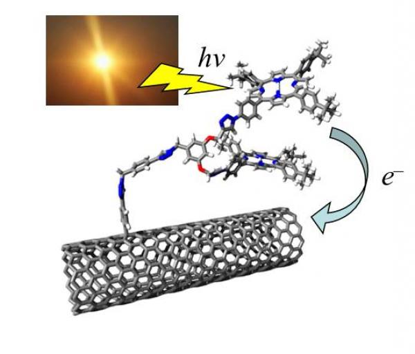
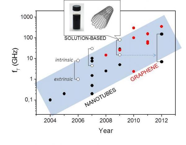
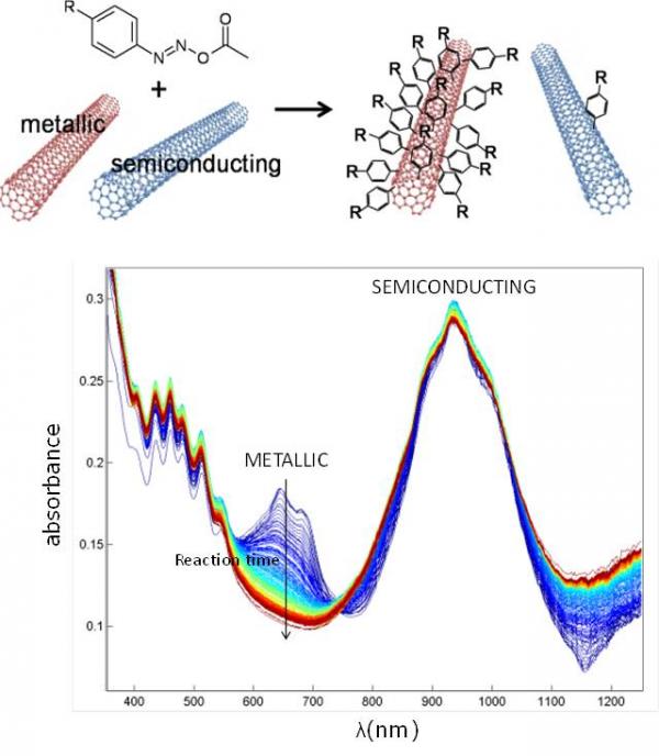
Single-wall carbon nanotubes (SWNTs) have exceptional electronic and optical properties. However, their synthesis produces a mixture of chiralities, some of which having a metallic character while the others are semiconducting. We studied in details the mechanism of diazonium coupling to SWNTs, identify the origin of the selectivity of this chemical reaction toward metallic nanotubes and use this understanding to optimize this selectivity to a point where the reacted material becomes relevant for applications of SWNTs as a semiconducting ink in organic electronics.
Photoelectron emission microscopy combined with intense photon sources is becoming a powerful analytical tool with both spatial resolution down to a few nanometers and spectroscopic capabilities. One can laterally resolve surface domains according to their magnetization, chemical composition, electronic structure, and even follow the dynamics of domain movement, catalysis or laser emissions using time resolved detection systems.
The lateral resolution varies from 1 mm to 10 nm, placing this technique at the heart of the mesoscopic physics, and corresponds to the need for spectroscopic information on the intermediate scale between atomic and macroscopic, so lacking in multiscale modeling. Furthermore, the possibility of studying nanostructured materials and objects on the 100 nm scale corresponds perfectly to the technologically interesting length scale in electronics, bioelectronics.
CEA has acquired the first production model of the NanoESCA (OMICRON GmbH), a spectromicroscope designed for high spatial and energy resolution over a wide range of photoelectron kinetic energies (Δx ~ 100 nm; ΔE ~ 100 meV). This is done thanks to the combination of a simple purely electrostatic lens column allowing accurate focus tracking over several hundred eV with the use of a double hemispherical analyzer, eliminating spherical aberrations usually present in energy analyzers for photoelectron microscopes.
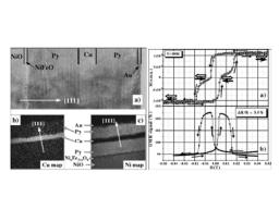
Fully epitaxial spin valve structures enable disentangling individual parameters contributing to the exchange bias effect. Antiferromagnetic oxides are used to pin the magnetization of ferromagnetic metal layers. The preparation of fully epitaxial spin valves elaborated on NiO(111) single crystals by means of molecular beam epitaxy techniques, with permalloy (Ni80Fe20), Co, Co30Fe70, and Co/Co30Fe70 as pinned layers, were investigated by surface X-ray diffraction, electron transmission microscopy, atomic force microscopy, vibrating sample magnetometry and electron transport measurements. [1] The best results were obtained with permalloy (Py) being the pinned layer with a giant magnetoresistance (GMR) of 3.5% at room temperature. The detrimental effect of an inter-diffusion at the ferromagnet/NiO interface has been evidenced. A 0.8-nm-thick CoFe layer has been found as being an excellent and efficient wetting layer for Co. These model structures allowed a decoupled investigation of the different parameters although the nearly isotropic GMR response of these structures makes them also interesting devices. The microstructure and the exchange coupling were investigated together. At room temperature, the major effect of the exchange coupling on single crystalline NiO(111) substrates is an inversely proportional behaviour of the coercive field of the pinned layer with respect to the ferromagnetic layer thickness. The effect is understood as resulting from the energy losses in the antiferromagnet because of irreversible losses in the antiferromagnetic order.
The growth mode and interface reactivity of the prototypical Co/α-Fe2O3(0001)/Pt(111)ferromagnet/antiferromagnet interface has been considered more recently using oxygen plasma assisted grown hematite epitaxial layers. [2] The chemical composition of the interface has been determined by means of X-ray absorption spectroscopy, X-ray magnetic circular and linear dichroism and the morphology of the growing Co film by scanning tunneling microscopy. We could evidence a 3D growth mode and the presence of a spatially limited reactive interface that spreads to approximately one atomic layer on either side of the interface. Kinetic limitations due to the diffusion of O in Co are most likely responsible for the very limited interfacial diffusion. This area is chemically composed by a layer of reduced hematite and a layer of oxidized cobalt. In the early stages of growth the interface composition is found to be dependent on the Co coverage and reaches quickly a steady state of fixed composition. At 2.5 nm the Co islands coalesce. X-PEEM magnetic domain imaging revealed a strong exchange coupling across the interface.
REFERENCES :
[1] C. Mocuta et al., Phys. Rev. B 68, 014416 (2003)
[2] O. Bezencenet et al., Surf. Sci. (2007)
Une communauté de chercheurs en spintronique
De nombreux travaux sont réalisés à l'IRAMIS dans le domaine de la spintronique, spécialité récemment mise à l'honneur avec le Prix Nobel décerné à Albert Fert (Dir. scientifique de l’UMR CNRS/Thales - Université Paris-Sud 11) et Peter Grünberg (Centre de recherches de Jülich, Allemagne).
Depuis plus de quinze ans, des collaborations croisées entre les chercheurs de l'IRAMIS (CEA), de l’Institut d’Electronique Fondamentale (CNRS), du Laboratoire de Physique des Solides (CNRS) et de l’unité mixte CNRS-Thales ont tissé de nombreux liens. Ainsi, en 1997, une collaboration entre l’unité d’Albert Fert et des chercheurs du CEA produisait un record mondial de magnétorésistance tunnel (TMR) dans des matériaux demi-métalliques. D’autres collaborations ont suivi… et aujourd'hui elles portent par exemple sur :
(Voir aussi la page thématique : "Nanomagnétisme et spintronique")
Matériaux multiferroïques [1]
Les matériaux « multiferroïques » (comme l’oxyde de bismuth et de fer : BiFeO3) se définissent par le cumul de propriétés électriques et magnétiques habituellement disjointes [2]. En couplant ces propriétés dans des multicouches ultra-minces, il est possible de contrôler un état magnétique grâce à un champ électrique et non plus seulement grâce à un courant. C’est une avancée décisive parce qu’à la différence d’un courant, un champ électrique (en pratique, une tension électrique) ne provoque aucun échauffement. Ces matériaux pourraient être utilisés avantageusement dans des mémoires MRAM, non volatiles et peu gourmandes en énergie.

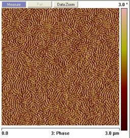
The majority of electronic devices is designed on silicon wafers where the architecture of the different layers is made through lithographic techniques and resin masks. To cope with the desired increase in performance (Moore’s law), the structure size has to be reduced in order to integrate a larger number of components. Lithographic techniques then reach limits (pure top-down process). To reach a resolution of order 22 nm (the next ITRS node) new methods like e-beam etching are discussed but constraints and costs are huge. Alternative solutions can be found thanks to molecular self-assembly (bottom-up processes). Among them, block copolymer self-assembly is attractive: block copolymers are made of different A and B monomers linked by covalent bonds. At low temperatures they form ordered structures (micro-phase separation) of domains at the desired scale (sone tens of nanometers, tunable with the molecular mass of the polymer). To use this phase separation, it is requested to orient in a controlled manner the obtained structures when they are under the form of a thin film on a wafer. A new convenient method was designed for providing a first perpendicular orientation which however presents lots of defects (Fig.)[1].
[1] Liu P.H., P. Thébault, P. Guenoun, J. Daillant, Macromolecules, 42, 9609 (2009)
Please contact: Patrick.Guenoun@cea.fr
http://iramis.cea.fr/Pisp/patrick.guenoun/index.html
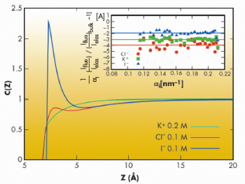
Les effets ioniques dits spécifiques regroupent tous les effets certes d'origine électrostatique mais qui ne peuvent être expliqués par la seule valence des ions. Ainsi, Cl- et Br- ou K+ et Na+ peuvent avoir des effets dramatiquement différents sur la structure et la stabilité colloïdale en biologie, en physicochimie organique et minérale, dans l'environnement… Au-delà du classement phénoménologique de Hofmeister (1888), la compréhension fine de ces effets a fait un bond en avant ces dernières années, d'une part grâce à des techniques modernes de mesure de profils ioniques aux interfaces, d'autre part grâce aux développements théoriques et numériques qui font intervenir le concept de polarisabilité ionique. Depuis quelques années, le LIONS développe ces deux facettes, en particulier depuis fin 2006 dans le cadre du projet ANR 066BLAN-0276 "Spécificité ionique dans les systèmes colloïdaux et aux interfaces" (SISCI). Il s'agit de comprendre les couplages électrostatiques à courte distance (en dessous du nm) entre ions, molécules de solvant et matériaux à la fois en solution et au voisinage des interfaces.
Du point de vue expérimental, le LIONS a développé différentes techniques utilisant la brillance des synchrotrons afin de mesurer le nombre et la distribution spatiale des ions adsorbés aux interfaces: fluorescence X sous incidence rasante à l'interface eau/air ([1], figure 1, stage post-doctoral Viswanath Padmanabhan), ondes stationnaires à l'inrerface liquide/solide.
See the PhD position opened in 2014 about this subject
http://iramis.cea.fr/Pisp/patrick.guenoun/Greenmembranes.html
Phase separation of polymer solution, induced by an abrupt quench in temperature, leads to a macroscopic segregation of phases. The growth of the phases and the associated structures when time elapses since the beginning of the quench are still badly known for hydrosoluble polymer solutions. However phase separation processes are widely used in manufacturing polymeric material like gels or membranes
Mastering the temperature-induced morphology and porosity needs to fully understand how phase separation proceeds and to establish the relationship between phase separation mechanism and processing parameters during material preparation.
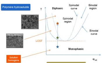
For some years, we studied a neutral hydrosoluble polymer the poly(N-isopropylacrylamide) or PNIPAM. When aqueous solutions of this polymer are heated above a temperature of the order of 42°C, a phase separation occurs. However the PNIPAM does not follow the macroscopic phase separation. The phase separation stops at a stage where it leads to porous stable PNIPAM colloids (spheres having size of the order of 200 nm). A probable mechanism, consistent with the colloidal size variation with temperature, is that the phase separation is blocked at an early stage (Cahn regime of spinodal decomposition) by adsorption of residual ions at the grain’s surface.
Our ultimate goal is now to make membrane manufacturing safer and more economic on atoms by using modified natural polymers like cellulose ethers and water as the only solvent, thus lowering wastes and recycling. Consolidation of the film structure is carried out by cross-linking. On the basis of their relevance for membrane preparation and accessible phase boundaries, the biocompatible and neutral polymer selected is the hydroxypropyl cellulose (HPC). Aqueous hydroxypropyl cellulose is a fascinating system which exhibits an isotropic phase in dilute solutions, but forms an ordered liquid crystalline phase with cholesteric structure in concentrated solutions at room temperature. As PNIPAM the HPC has a lower critical solution temperature (LCST) close to room temperature and it undergoes reversible phase separation upon heating. Phase diagrams of HPC solutions will be established and bulk phase separations will be studied to determine what kind of bicontinuous phases are obtained as a function of the temperature, concentration and time. The characterization will be done in a large concentration range in order to change the topologies of the predominant phase (in volume) rich in polymer or in solvent. Phase diagrams and bulk phase separations of HPC solutions will be studied by varying the physico-chemical parameters: degree of polymerization and polydispersity.
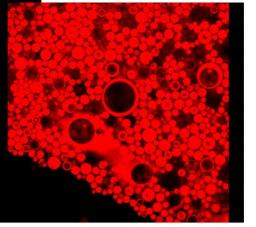
The boundaries of the phase diagrams (cloud points) are determined by turbidity measurements. The morphologies and the growth laws with time of phases are established using scattering techniques (Neutron, Light, XRay), Diffusive Wave Spectroscopy, Optical and Confocal Microscopies (see Figure 1).
Quantitative models will be proposed to rationalize the phase separation processes in order to provide roadmaps for membrane manufacturing.
Quelle que soit la nature des applications envisagées (émulsions, protection de colloïdes, lubrification...) les propriétés des films interfaciaux dépendent intrinsèquement de leur structure et de leur réponse à une contrainte externe. Cette réponse se traduit par des changements structuraux et par des comportements mécaniques que nous avons étudiés, le plus souvent en lien avec des études présentées ailleurs dans ce document.
Notamment, les études sur les films de polymères présentées ici permettent d'éclairer le comportement de ces mêmes polymères en volume lorsque l’auto-assemblage génère un grand nombre d’interfaces comme dans les émulsions ou micro-émulsions (voir "Stabilisation d'émulsions"). La liberté de fixer l'aire par chaîne en surface donne accès à une grande richesse de comportements qui permettent de tester la théorie et de mettre en perspective les résultats obtenus en volume. Ces travaux se sont appuyés sur des développements expérimentaux novateurs, souvent auprès de grands instruments, et sur des synthèses originales réalisées par J.W. Mays (U. Tennessee). De même, il y a un lien étroit entre les travaux sur les bicouches lipidiques présentés ici et les développements ambitieux utilisant des cyclodextrines modifiées (voir "Cyclodextrines modifiées: de l'inclusion à la fonctionnalisation des membranes").
Une très grande richesse de comportements est également observée dans la manière dont des interfaces décorées par des polymères ou des amphiphiles vont réagir à une contrainte, par exemple une réduction de l'aire interfaciale. S'il est encore trop tôt pour dégager une description générale de ces phénomènes, des comportements inattendus ont pu être observés et expliqués, comme l'apparition de réseaux réguliers de stries à l'interface, ou l'apparition de fluctuations géantes pouvant être expliquées par une tension de surface effective très faible.

Ecotoxicological impacts of Nanoparticles
Thanks to their particular or enhanced properties due to their size, the nanomaterials (dimension < 100 nm) are widely used in many industrial applications (daily care products, nanostructured materials...). However, their growing use together with the belief of easy dispersability frightens because of their uncertain impact on humans and environment. Our work is dedicated to a deep understanding of the physicochemical and biological interactions between cellular models such as: Synechocystis (cyanobacteria essential for the biosphere) or Escherichia coli (bacteria of mammalian intestines) with nanoparticles (CeO2, TiO2, Ag0, Au).
The particular behavior of nanoparticles (agregation, dissolution, adsorption...) requires a different and highly multidisciplinary approach compared to the study involving classic chemical compounds. Indeed, we showed that the physicochemical parameters (stability, aggregation, dissolution and surface chemistry) of nanoparticles in the contact medium, strongly influence the toxicity. Furthermore, the physicochemical interactions (flocculation, adsorption, redox mechanisms) are linked to the biological model; for exemple, the presence of exopolysaccharides as natural barrier between the cell wall and the nanoparticles. Moreover, the composition of the nanoparticles dispersion medium (particularly the pH) has a major influence on the toxicity.
Many of these research activities are carried in the framework of the international Consortium for the Environemental Impact of Nanotechnologies (iCEINT). The LIONS is also leading a CNano Project of the region Ile de France, regarding the interaction between nanoparticles and bacteria in the Seine River. The project is at the frontier between physics and chemistry of NPs, biology and ecotoxicology.
Sous l’effet de la compression, la monocouche subit une transition de phase, qui fait apparaître une direction cristalline supplémentaire, perpendiculairement aux feuillets β. La transition du cristal peptidique 1D à un cristal 2D est décrite en termes de formation de structures hiérarchiques (cf. Fig.1) : les "cristaux 1D" initiaux sont des "nanobandes" (feuillets β individuels) de 100 Å de large et ~ 100 nm de long qui s’associent latéralement pour former des "cristaux 2D" appelés nanodomaines, dix fois supérieurs en taille mais de même rapport d’aspect. [3-4]
Ces structures pourraient s’apparenter aux structures d’interface, différentes des fibres de volume,formées par certaines molécules amyloïdes naturelles adsorbées à des interfaces biologiques. A forte compression, les nanodomaines peptidiques constituent un film quasi-continu polycristallin qui, après dépôt sur un substrat solide, servira de moule organique dans de prochaines expériences de minéralisation. Nous souhaitons par ailleurs préciser la structure de ces cristaux peptidiques à une échelle quasi-atomique en étendant au cas des films d’oligopeptides une technique de cristallographie 2D développée récemment au LIONS (cf. "Structure et élasticité des films amphiphiles et brosses de polymères").
La stabilisation d’émulsions (hors d’équilibre) ou de microémulsions (à l’équilibre thermodynamique) est un processus industriel d’importance mais qui suscite toujours de nouveaux développements fondamentaux liés notamment à la recherche de tensioactifs plus performants ou à une meilleure compréhension des mécanismes d’inversion.
Les tensioactifs ‘‘sophistiqués’’ que nous avons utilisé ont vu le jour grâce à des synthèses chimiques originales comme des copolymères réalisés par polymérisation radicalaire vivante à l’ESPCI (PS-PDAEMA) ou par polymérisation anionique sous vide (laboratoire de J. Mays, Knoxville) ou encore des nanoparticules greffées avec des ligands idoines. Ce sont souvent des objets stimulables qui s’intègrent dans un objectif général de modulation des propriétés en fonction de paramètres extérieurs facilement accessibles (température, pH, ajout de sel...). Ces mêmes molécules ou nanoparticules ont bénéficié pour la plupart d’études en film unique présentées ici ou dans la fiche ‘‘Interfaces’’, ce qui permet d’enrichir la compréhension des émulsions en tant qu’auto-assemblage de films flexibles. Enfin, des techniques alliant le suivi, parfois cinétique, dans l’espace direct et dans l’espace réciproque ont été utilisées.
Des micro-émulsions peuvent être stabilisées à l’aide de copolymères biséquencés neutres-chargés qui jouent alors le rôle de tensioactifs dont les deux parties hydrophiles et hydrophobes sont aisément modulables par construction ou ajout de sel. C’est notamment possible en utilisant des copolymères seuls qui forment des micelles gonflées [1] par un bon solvant du coeur hydrophobe et ce d’autant plus que les deux séquences sont plus équilibrées. Par contre, si l’on se sert des mêmes copolymères comme additif à une microémulsion bicontinue stabilisée par des tensioactifs courts de type CiEj, on obtient alors un ‘‘boosting’’ de la microémulsion. Comme montré Figure 3, l’ajout de copolymère permet à la phase microémulsion de gonfler en incorporant de l’huile et eau supplémentaires et l’utilisation de copolymères chargés nous a permis d’atteindre des facteurs de boosting deux fois plus grands que ceux rapportés jusqu’alors dans la littérature comme en témoigne la teinte blanche de la phase du milieu, au centre de la Figure 1. En fonction de la température, on peut varier le gonflement qui est plus ou moins important et symétrique. Une explication possible est liée à l’accroissement de la rigidité de courbure du film par l’ajout de copolymère pour une courbure spontanée du film proche de zéro.

General objectives
We aim at understanding the microscopic mechanisms controlling the formation of nanoparticles. Different types of NPs are studied in the LIONS and among them gold NPs have already been the subject of several achieved PhD (B. Abécassis and F. Hubert) and on-going in-house studies (J. Han) and collaborative ones (S. Gomez from the Univiersity og Vigo). Our general approach is to consider that quantitative kinetic measurements of the size, size distribution and number of NPs with time together with the gold atoms speciation in solution (Au(III), Au(I) and Au-0)) can serve as a basis to develop a better understanding of the mechanisms involved.
The nucleation and growth mechanism of gold nanoparticles are followed at the millisecond time scale using different in situ techniques. Three different systems are presently studied : nanospheres in toluene (NST), nanorods in water (GNR) and nanospheres in water (NSW). The time scale range from one second (NSW) to 600 seconds (GNR). The sample environment are adapted to the time-scale involved. They are (i) a stopped for the most rapid synthesis, (ii) an home-made millifuidic set-up for water rapid systems and (iii) a simple flow through cell for the GNRs.
Three main synchrotron based techniques are used, SAXS WAXS and XANES both with a high time resolution. ESRF (ID02) and SOLEIL (SWING and ODE) have been intensively used to acquire a unique set of data during the nucleation and growth of these objects.
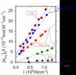
The ionization of the DNA single and double helices (dA)20, (dT)20, dAdT)10.(dAdT)10 and (dA)20.(dT)20, induced by nanosecond pulses at 266 nm, is studied by time-resolved absorption spectroscopy. The variation of the hydrated electron concentration with the absorbed laser intensity shows that, in addition to two photon ionization, one photon ionization takes place for (dAdT)10(dAdT)10, (dA)20(dT)20 and (dA)20 but not for (dT)20. The spectra of all adenine containing oligomers at the microsecond time-scale correspond to the adenine deprotonated radical formed in concentrations comparable to that of the hydrated electron. The quantum yield for one photon ionization of the oligomers (ca. 10-3) is higher by at least one order of magnitude than that of dAMP showing clearly that organization of the bases in single and double helices leads to an important lowering of the ionization potential. The propensity of (dAdT)10(dAdT)10, containing alternating adenine – thymine sequences, to undergo one photon ionization is lower than that of (dA)20(dT)20 and (dA)20, containing adenine runs. Pairing of the (dA)20 with the complementary strand leads to a decrease of quantum yield for one photon ionization by about a factor two.
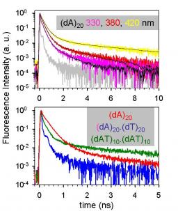
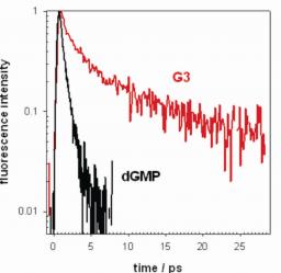
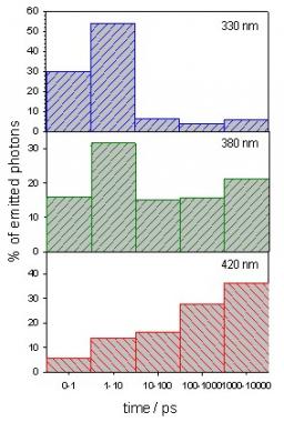
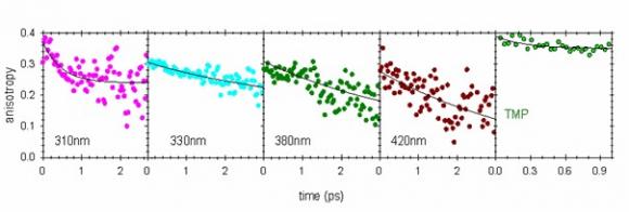
Absorption of ultraviolet light by DNA is known to lead to carcinogenic mutations, but the processes between photon absorption and the photochemical reactions are poorly understood. In their study of the excited-stated dynamics of model DNA helices using femtosecond transient absorption spectroscopy1, Crespo-Hernández et al. observe that the picosecond component of the transient signals recorded for the adenine–thymine oligonucleotide (dA)18•(dT)18 is close to that for (dA)18, but quite different from that for (dAdT)9•(dAdT)9; from this observation, they conclude that excimer formation limits excitation energy to one strand at a time. Here we use timeresolved fluorescence spectroscopy to probe the excited-state dynamics, which reveals the complexity of these systems and indicates that the interpretation of Crespo-Hernández et al. is an oversimplification. We also comment on the pertinence of separating base stacking and base pairing in excited-state dynamics of double helices and question the authors’ assignment of the long-lived signal component found for (dA)18•(dT)18 to adenine excimers. Figure 1a shows the fluorescence decays of (dA)20 at three different wavelengths. Combining these results with our previous measurements obtained for (dA)20 by femtosecond fluorescence upconversion2, at least five exponentials are needed to fit the decays over the 100 femtoseconds to 20 nanoseconds time range. A crucial point is that all time constants vary strongly with the emission wavelength. The same effect is encountered for (dAdT)10•(dAdT)10 and has been reported previously3 for poly(dA)•poly(dT).
The investigation of model DNA helices using time-resolved absorption and fluorescence spectroscopy with UVB/UVC excitation knows currently an increasing interest due, in particular, to the use of femtosecond spectroscopy. The study of such complex and fragile systems presents specific difficulties which are not encountered in the experiments performed on the monomeric DNA units. They are related both to the quality of the DNA helices and their sensitivity towards UV radiation. The present paper tackles some of these problems (pitfalls) and describes experimental protocols developed in our laboratory in order to overcome them (tricks). We focus on experiments carried out by fluorescence upconversion spectroscopy, time-correlated single photon counting and nanosecond flash photolysis. We illustrate our experience with examples obtained for double helices containing only adenine-thymine base pairs. We consider this report of pitfalls and tricks, which is far from being complete, as a first step towards a codification of rules for time-resolved studies with model DNA helices. This codification has to be established by common agreement of the various groups working in the field.
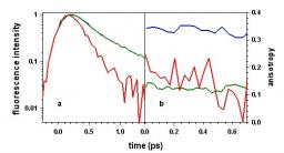
The DNA double helix poly(dGdC).poly(dGdC) is studied by fluorescence upconversion spectroscopy with femtosecond resolution. It is shown that the excited state relaxation of the duplex is faster than that of the monomeric components dGMP and dCMP. This contrasts with the behavior of duplexes composed exclusively of adenine-thymine base pairs, for which an overall lengthening of the fluorescence lifetimes with respect to that of an equimolar mixture of dAMP and TMP was reported previously. Despite the difference in the excited state deactivation rate between the two types of duplexes, the signature of ultrafast energy transfer is present in both of them. It is attested by the decrease of fluorescence anisotropy decay of the duplexes on the sub-picosecond time scale, where molecular motions are inhibited, and is corroborated by the fact that their steady-state fluorescence spectra do not change with the excitation wavelength. Energy transfer involves excited states delocalized over at least two bases, whose existence is revealed by the UV absorption spectrum of the duplex, clearly different from that of an equimolar spectrum of dGMP and dCMP.
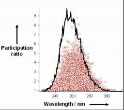
The present communication deals with the singlet excited states of the model DNA duplex (dA)10.(dT)10. Calculations are performed in the frame of the exciton theory, combining quantum chemistry methods and molecular dynamics simulations. The monomer transition energies are simulated by Gaussian functions resembling the absorption bands of nucleosides in aqueous solutions. Most of the excited states are found to be delocalized over at least two bases and result from mixing of different monomer states. Their properties are only weakly affected by conformational changes of the double helix. On average, the highest oscillator strength is carried by the upper eigenstates. The duplex absorption spectra are shifted a few nanometers to higher energies with respect to the spectra of non-interacting monomers. The states with larger spatial extent are located close to the maximum of the absorption spectrum.
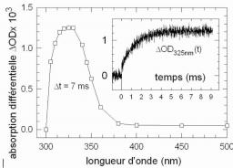
The formation of thymine dimers in the single stranded oligonucleotide (dT)20 is studied at room temperature by laser flash photolysis using 266 nm excitation. It is shown that the (6-4) adduct is formed within 4 ms via a reactive intermediate. The formation of cyclobutane dimers is faster than 200 ns. The overall quantum yield for the (6-4) formation is (3.7 +/- 0.3)x10-3 and that of the cyclobutane dimers (2.8 +/- 0.2)x10-2. No triplet absorption is detected showing that either the intersystem crossing yield decreases by one order of magnitude upon oligomerization (<1.4x10⁻³) or the triplet state reacts with unit efficiency in less than 200 ns to yield cyclobutane dimers.
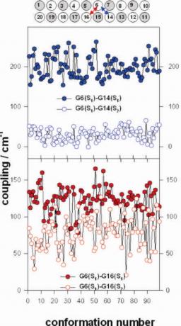
The present communication deals with the excited states of the alternating DNA oligomer (dCdG)5.(dCdG)5 which correspond to the UV absorption band around 260 nm. Their properties are studied in the frame of the exciton theory, combining molecular dynamics simulations and quantum chemistry data. It is shown that the dipolar coupling undergoes important variations with the site and the helix geometry. In contrast, the energy of the monomer transitions within the double helix is not sensitive to the local environment. It is thus considered to be distributed over Gaussian curves whose maximum and width are derived from the experimental absorption spectra of nucleosides in aqueous solution. The influence of the spectral width on the excited states delocalization and the absorption spectra is much stronger than that of the oligomer plasticity. About half of the excited states are delocalized over at least two bases. Many of them result from mixing of different monomer states and extend on both strands. The trends found in the simulated spectra, when going from non-interacting monomers to the duplex, are in agreement with experimental observations. Conformational changes enhance the diversity of the states which can be populated upon excitation at a given energy. The states with larger spatial extent are located close to the maximum of the absorption spectrum.
The first fluorescence investigation of DNA oligomers performed with femtosecond resolution revealed that organisation of nucleotides into single strands, and further into double strands, make the fluorescence decays slower and slower. This is important because a longer lifetime increases the reactivity of the excited states and, thus, the probability to induce damages of the double helix

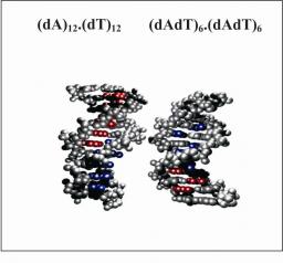
The present communication addresses the question of the magnitude of dipolar coupling between the lowest electronic transition moments of the DNA nucleosides and its relevance for Frenkel exciton states in double helices. The transition energies and moments of the nucleosides are determined from absorption spectra recorded for dilute water solutions. Dipolar interactions are computed for some typical nucleoside dimers according to the atomic transition charge distribution model. The properties of the exciton states of two particular double helices, (dA)20.(dT)20 and (dAdT)10.(dAdT)10, are calculated considering three closely-lying molecular electronic transitions (S1 and S2 for adenosine, S1 for thymidine). It is shown that (i) the oscillator strength is distributed over a small number of eigenstates, (ii) important mixing of the three monomer electronic transitions may occur, (iii) all eigenstates are spatially delocalised over the whole length of the double helix and (iv) the extent of exciton states over the two strands depends on the base sequence.
The present communication deals with the photophysical properties of triguanosine diphosphate in aqueous solutions which are compared with those of the 2’-deoxyguanosine monophosphate. They are studied by steady-state absorption and fluorescence spectroscopy as well as by time-resolved fluorescence spectroscopy with femtosecond resolution. The temperature, salt and concentration dependence of the absorption and fluorescence spectra reveal that association of the trimers takes place. The resulting aggregates could correspond to a tetraplex structure. The aggregate fluorescence quantum yield is higher and the fluorescence lifetime much longer than those of the monomer. These results show the cooperativity between guanosine residues which may manifest itself via self-solvation, hydrogen bonding and/or delocalization of the excitation.
Absorption of UV radiation by DNA bases is known to induce carcinogenic mutations. The lesion distribution depends on the sequence around the hotspots suggesting cooperativity between bases. Here we show that such cooperativity may intervene at the very first step of a cascade of events by formation of Franck-Condon states delocalized over several bases and subsequent energy transfer faster than 100 fs. Our study focuses on the double helix poly(dA).poly(dT), whose fluorescence, induced by femtosecond pulses at 267 nm, is probed by fluorescence upconversion and time-correlated single photon counting, over a large time domain (100 fs – 100 ns). The time-resolved fluorescence decays and fluorescence decays are discussed in relation with the steady-state absorption and fluorescence spectra in the frame of exciton theory.
The present communication examines how the dynamics of the double helix affects the Frenkel excitons corresponding to the low-energy absorption band of DNA. Two types of oligomers, (dA)n.(dT)n and (dAdT)n/2.(dAdT)n/2, are studied theoretically, in the frame of the exciton theory in combination with quantum chemical calculations. The properties of the exciton states (energy, oscillator strength, degree of delocalization, “anisotropy”…) found for canonical B-DNA geometries are compared to those obtained for conformations extracted from molecular dynamics simulations. It is shown that although structural fluctuations reduce both the mixing between different monomer transitions and the spatial extent of the eigenstates, excitations still remain delocalized over several bases. The presence of alternating base sequences makes the eigenstates of the double-stranded oligomers more sensitive to disorder. All these effects arise from a variation of the coupling terms, the diagonal energy being only slightly altered by the structural fluctuations. The experimental absorption spectra presented here corroborate the theoretical results according to which the absorption of (dA)n.(dT)n is centered at higher energies than that of (dAdT)n/2.(dAdT)n/2. Finally, it is shown that, in contrast to what is commonly admitted, the formation of collective excited states in double-stranded oligomers is not expected to induce large spectral shifts with respect to a homogeneous mixture of monomers.
The study of excited states and energy transfer in DNA double helices has recently gained new interest connected to the development of computational techniques and that of femtosecond spectroscopy. The present article points out contentious questions regarding the nature of the excited states and the occurrence of energy transfer and shows how they are currently approached. Using as example the polymer poly(dA).poly(dT), composed of about 2000 adenine-thymine pairs, a model is proposed on the basis of time-resolved measurements (fluorescence decays, fluorescence anisotropy decays and fluorescence spectra, obtained with femtosecond resolution), associated to steady-state spectra. According to this qualitative model, excitation at 267 nm populates excited states that are delocalized over a few bases (excitons). Ultrafast internal conversion directs the excited state population to the lower part of the exciton band giving rise to fluorescence. Questions needing further investigations, both theoretical and experimental, are underlined with particular emphasis on delicate points related to the complexity and the plasticity of these systems.
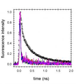
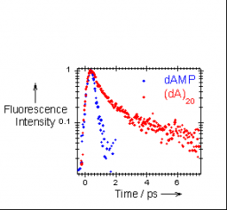
The fluorescence of the DNA double stranded oligomer (dA)20.(dT)20 is studied at room temperature by fluorescence upconversion at times shorter than 10 ps. The profile of the upconversion spectra is similar to that of the steady-state fluorescence spectrum, showing that the major part of the photons is emitted within the probed time-scale. At all the probed wavelengths, the fluorescence decays are slower than those of the monomeric chromophores dAMP and TMP. The fluorescence anisotropy decays show strong wavelength dependence. These data allows us to conclude that energy transfer takes place in this double helix and that this process involves exciton states. The spectral and dynamical properties of the oligomer are compared to those of the polymer poly(dA).poly(dT), composed of about 2000 base pairs, reported previously. The oligomer absorption spectrum is characterized by a smaller hypsochromic shift and weaker hypochromism compared to the polymer. Moreover, the fluorescence decays of (dA)20.(dT)20 are a twice as fast as those of poly(dA).poly(dT) and its fluorescence anisotropy decays more slowly. These differences are the fingerprints of a larger delocalization of the excited states induced by an increase in the size of the duplex.
Fluorescence of the DNA double helix (dA)20·(dT)20 studied by femtosecond spectroscopy : effect of the duplex size on the properties of the excited states
D. Onidas, T. Gustavsson
Cette thématique phare du groupe vise à collecter des données structurales précises en phase gazeuse afin d’étalonner la modélisation des interactions moléculaires contrôlant la structure des biomolécules.
Les atouts du groupe sont ici la maîtrise d'expériences en phase gazeuse, (désorption par laser, jet supersonique et méthodes de spectroscopie optiques laser à plusieurs couleurs, UV et IR) et le développement poussé de l’interface entre expérience et théorie (modélisation des structures et spectres IR par chimie quantique, modélisation des processus en détente supersonique, validation des nouveaux outils théoriques pour la description des systèmes complexes).
Il s’agit ici d’aborder une question cruciale pour les systèmes biologiques, à savoir celle du lien entre structure (conformation, environnement), et dynamique (réactivité, relaxation).
Cette thématique est en prise directe avec les propriétés des protéines, vis-à-vis du devenir de l’excitation électronique consécutive à l’absorption d’un photon dans le proche UV. Les états excités de longue durée de vie laissent le temps à des réactions chimiques de se produire, conduisant à des dommages structurels susceptibles d’affecter la fonction biologique du système. Or, de manière générale, les molécules du vivant présentent des états excités de courte durée de vie, court-circuitant ainsi les phénomènes dommageables : ce sont ces phénomènes élémentaires contrôlant la durée de vie de l’état excité qui sont ciblés dans ces études. Il s’agit ici de :
- caractériser expérimentalement les durées de vie en fonction des espèces étudiées (expériences de type pompe-sonde), ainsi que la cascade d’états formés,
- modéliser les processus mis en jeu, notamment le rôle joué, pour la relaxation, par les intersections coniques avec des états excités de plus basse énergie.
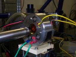
Experiments using diamond anvil cells (DAC) do not allow a very precise determination of the pressure and temperature parameters, more especially in the case of hydrothermal DAC such as Bassett type at high temperatures.
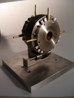
Therefore, we are working on a new kind of cell: the “intelligent” DAC (Burchard & al, Rev. Sci. Instr. 74, 2003, Figure 1) in collaboration with the Ruhr Universität Bochum, Germany.
In this cell, diamonds are modified in order to become pressure and temperature sensors. Therefore a piezoelectric structure is implanted below the diamond culets (Figure 2) using a particle accelerator (ARAMIS, CSNSM, IN2P3, Orsay, and Ruhr Universität Bochum, Germany).
Pressure and temperature calibrations are now under progress.
Micro-experimental studies require the use of diamond anvil cells, allowing observation and in situ characterization of the studied phase equilibrium using classical spectroscopy techniques (Infrared, Raman, X-Ray Fluorescence, X-Ray diffraction…).
In the Bassett’s cell, the sample is contained in a rhenium gasket compressed by two type I diamonds of 0.21 carat with 1 mm flats. The initial diameter of the borehole in the gasket is 0.5 mm. Heating is achieved by passing a high electrical current through the molybdenum wires around the tungsten carbide seats below the diamonds. During operation, the cell is flushed with an argon-hydrogen (2 %) mixture to prevent oxidation of the diamonds and the molybdenum heater. Temperature is measured to ± 2˚C by two type K thermocouples attached to the diamonds. The temperature in the sample chamber is calibrated against these thermocouples by measuring the melting points of NaNO3, CsCl, NaCl, sulfur and ice in the cell at room pressure.
Pressure medium in all experiments is water. Pressure at a given temperature is calculated from the equation of state of water. This is based on the observation that the sample cavity behaves as an isochoric system after several cycles of heating and cooling. Accordingly, the gasket is first filled with water and an air bubble to give the desired bulk density and cycled through the relevant temperature range several times, until the homogenization temperature of the fluid is virtually constant. After that the cell is loaded with the sample (ships of glass, crystals…), water (or aqueous solution) and an air bubble, then it is heated to the desired temperature. After cooling, the bulk density of the pressure medium is determined by measuring the homogenization temperature, if a bubble was present, or the melting point of ice, if there is no bubble. From this bulk density, the pressure is then calculated for the temperatures prevailing during the experiment.
Observation and measurements are performed in situ at high pressure (up to 2 GPa) and high temperature (up to 1100°C) through the diamonds along the compression axis.
See also : http://mplmac.geo.cornell.edu/HDAC/.
Les protéines, molécules indispensables à la vie des organismes vivants, sont des polymères synthétisés à partir de l’ADN des cellules. Leur fonction dépend de leur constitution mais aussi de leur structure. Ces molécules sont constituées de longues chaînes linéaires d'acides aminés (typiquement une centaine) ; c’est ce qu’on appelle la structure primaire des protéines. Si l’on réalise que 20 types différents d’acides aminés existent dans le vivant et que chacun d’eux peut présenter plusieurs structures différentes, on conçoit que cette stratégie ait donné lieu à une très grande diversité de structures et de fonctions. L’un des points clés contrôlant la structure des protéines est le processus de repliement conduisant le polymère linéaire à adopter une structure tridimensionnelle. La première phase du repliement conduit à la formation de structures-type, appelées structures secondaires, concernant un nombre restreint d’acides aminés de 2 à quelques dizaines, dont les plus connues sont les feuillets beta ou les hélices alpha. Le repliement se poursuit ensuite par l’agencement relatif des structures secondaires entre elles.
Comprendre et décrire le vivant, notamment avec les outils modernes de simulation numérique, nécessite donc d'appréhender ces organisations structurales et de connaître précisément les interactions physicochimiques qui les gouvernent. Une approche nouvelle, développée dans l’équipe "Biomolécules excitées" du SPAM/LFP à Saclay, consiste à s’intéresser à de petits segments de protéines - les chaînes peptidiques- et à les faire se replier sous l’action de leurs propres interactions intramoléculaires. En pratique, on porte ces peptides en phase gazeuse à haute température dans un état désordonné "dénaturé", par une technique de désorption par laser, puis, dans une détente supersonique, on les laisse se refroidir par collisions avec le gaz porteur. Le repliement obtenu est alors analysé a posteriori, avec toute la précision des spectroscopies laser les plus modernes (technique de double-résonance IR/UV). Le croisement des informations spectroscopiques électroniques et vibrationnelles permet alors de caractériser les minimas locaux du paysage conformationnel balayé, notamment au travers de leur réseau de liaisons hydrogène intramoléculaires.
Publications
Intramolecular chiral recognition in a jet-cooled short peptide chain: gamma-turn helicity probed by a neighbouring residue;
Gloaguen,E.; Pagliarulo, F.; Chin, W.; Brenner, V.; Piuzzi, F.; Tardivel, B.; Mons, M.
Phys. Chem. Chem. Phys. 2007, 9, 4491; Hot Paper, Invited paper in a Special Issue on the spectroscopic probes of Molecular recognition.
Spectroscopic Evidences for the Formation of Helical Structures in Gas Phase Short Peptide Chains;
Brenner, V.; Piuzzi, F.; Dimicoli, I.; Tardivel, B.; Mons, M.
J. Phys. Chem. A 2007, 111, 7347, Invited paper in a Special Issue dedicated to R.E. Miller.
Chirality-controlled formation of beta-turn secondary structures in short peptide chains : experiment vs. quantum chemistry;
Brenner, V.; Piuzzi, F.; Dimicoli, I.; Tardivel, B.; Mons, M.
Angew. Chem. Int. Ed. 2007, 46, 2463.
Chin, W.; Piuzzi, F.; Dimicoli, I.; Mons, M.
Phys. Chem. Chem. Phys. 2006 8, 1033 (Invited Paper)
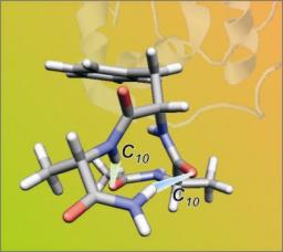
Gas phase formation of a 310-helix in a three-residue peptide chain: Role of side chain-backbone interactions as evidenced by IR-UV double resonance experiments;
Chin, W.; Piuzzi, F.; Dognon, J. P.; Dimicoli, I.; Tardivel, B.; Mons, M.
J. Am. Chem. Soc. 2005 127, 11900
Spectroscopic evidence for gas-phase formation of successive beta-turns in a three-residue peptide chain;
Chin, W.; Compagnon, I.; Dognon, J. P.; Canuel, C.; Piuzzi, F.; Dimicoli, I.; von Helden, G.; Meijer, G.; Mons, M.
J. Am. Chem. Soc. 2005 127, 1388
Intrinsic folding of small peptide chains: Spectroscopic evidence for the formation of beta-turns in the gas phase;
Chin, W.; Dognon, J. P.; Piuzzi, F.; Tardivel, B.; Dimicoli, I.; Mons, M.
J. Am. Chem. Soc. 2005 127, 707
Secondary structures of Val-Phe and Val-Tyr( Me) peptide chains in the gas phase: effect of the nature of the protecting groups;
Chin, W.; Dognon, J. P.; Piuzzi, F.; Dimicoli, I.; Mons, M.
Mol. Phys. 2005 103, 1579
Secondary structures of short peptide chains in the gas phase: Double resonance spectroscopy of protected dipeptides;
Chin, W.; Dognon, J. P.; Canuel, C.; Piuzzi, F.; Dimicoli, I.; Mons, M.; Compagnon, I.; von Helden, G.; Meijer, G.
J. Chem. Phys. 2005 122, 054317
Gas-phase models of gamma turns: Effect of side-chain/backbone interactions investigated by IR/UV spectroscopy and quantum chemistry;
Chin, W.; Piuzzi, F.; Dognon, J. P.; Dimicoli, I.; Mons, M.
J. Chem. Phys. 2005 123, 084301
The gas-phase dipeptide analogue acetyl-phenylalanyl-amide: A model for the study of side chain/backbone interactions in proteins;
Chin, W.; Mons, M.; Dognon, J. P.; Mirasol, R.; Chass, G.; Dimicoli, I.; Piuzzi, F.; Butz, P.; Tardivel, B.; Compagnon, I.; von Helden, G.; Meijer, G.
J. Phys. Chem. A 2005 109, 5281
Characterization of the conformational probability of N-acetyl-phenylalanyl-NH2 by RHF, DFT, and MP2 computation and AIM analyses, confirmed by jet-cooled infrared data;
Chass, G. A.; Mirasol, R. S.; Setiadi, D. H.; Tang, T. H.; Chin, W.; Mons, M.; Dimicoli, I.; Dognon, J. P.; Viskolcz, B.; Lovas, S.; Penke, B.; Csizmadia, I. G.
J. Phys. Chem. A 2005 109, 5289
Competition between local conformational preferences and secondary structures in gas-phase model tripeptides as revealed by laser spectroscopy and theoretical chemistry;
Chin, W.; Mons, M.; Dognon, J.-P.; Piuzzi, F.; Tardivel, B.; Dimicoli, I.
Phys. Chem. Chem. Phys. 2004 6, 2700 (PCCP Hot Paper)
A simple laser vaporization source for thermally fragile molecules coupled to a supersonic expansion: application to the spectroscopy of tryptophan;
Piuzzi, F.; Dimicoli, I.; Mons, M.; Tardivel, B.; Zhao, Q.
Chem. Phys. Lett. 2000 320, 282
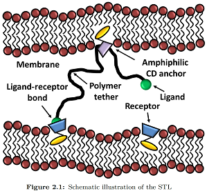
Recently, it has been theoretically proposed that polyrotaxanes, the inclusion complexes formed between a polymer and a ring-like molecule, could lead to a new class of grafted polymer layers whereby the attachment point between the chain and the substrate would allow the chains to slide freely, while being prevented from escaping by a capping group at each chain-end chosen to allow biorecognition. This form of chain attachment to a surface leads to a new class of polymer interfacial structure, that we name Sliding Anchored Polymers, or SAPs, with many new interesting potential features from the point of view of both equilibrium and dynamic layer properties.
Ecotoxicological impacts of Nanoparticles
Thanks to their particular or enhanced properties due to their size, the nanomaterials (dimension < 100 nm) are widely used in many industrial applications (daily care products, nanostructured materials...). However, their growing use together with the belief of easy dispersability frightens because of their uncertain impact on humans and environment. Our work is dedicated to a deep understanding of the physicochemical and biological interactions between cellular models such as: Synechocystis (cyanobacteria essential for the biosphere) or Escherichia coli (bacteria of mammalian intestines) with nanoparticles (CeO2, TiO2, Ag0, Au).
The particular behavior of nanoparticles (agregation, dissolution, adsorption...) requires a different and highly multidisciplinary approach compared to the study involving classic chemical compounds. Indeed, we showed that the physicochemical parameters (stability, aggregation, dissolution and surface chemistry) of nanoparticles in the contact medium, strongly influence the toxicity. Furthermore, the physicochemical interactions (flocculation, adsorption, redox mechanisms) are linked to the biological model; for exemple, the presence of exopolysaccharides as natural barrier between the cell wall and the nanoparticles. Moreover, the composition of the nanoparticles dispersion medium (particularly the pH) has a major influence on the toxicity.
Many of these research activities are carried in the framework of the international Consortium for the Environemental Impact of Nanotechnologies (iCEINT). The LIONS is also leading a CNano Project of the region Ile de France, regarding the interaction between nanoparticles and bacteria in the Seine River. The project is at the frontier between physics and chemistry of NPs, biology and ecotoxicology.
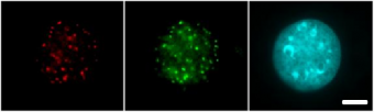
Nous avons réalisé l'étude de la réponse de cellules ostéoblastiques (MC3T3) à des irradiations contrôlées et ciblées à l'aide du dispositif d'irradiation ion par ion. Cette étude apporte un nouveau regard sur l'effet de voisinage (plus connu sous l'appellation bystander effect), dont l'origine est ici double: signalisation membranaire et mitochondriale. Jusqu'à présent, l'effet bystander était en général évoqué pour expliquer l'apparition d'effets délétères dans des cellules non directement ciblées. Ici, outre cette observation, nous avons également fait apparaitre un effet d'amplification dans les cellules cibles elles-mêmes: ainsi, pour une dose par cellule identique, la réponse à l'irradiation (évaluée par différents marqueurs de l'irradiation comme l'apparition de micronoyaux) est considérablement augmentée quand les cellules voisines sont elles-mêmes irradiées. Il faut donc dans notre cas élargir ainsi le concept de l'effet bystander également aux cellules cibles.
|
Merci de lciquer ici pour accéder au nouveau site web Ultrafast Nanophotonics |

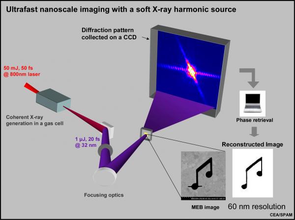
Imager des objets individuels avec une résolution spatiale de quelques nanomètres et une résolution temporelle de quelques femtosecondes revêt une importance fondamentale dans de nombreux domaines de la science et constitue aujourd’hui un défi fascinant. Les progrès de la diffraction cohérente de rayon X ont démontré le très grand potentiel de cette technique pour imager des structures nanométriques uniques et non périodiques[Miao, Nature 400 (1999); Pfeifer et al., Nature 442 (2006)]. L’imagerie par diffraction X cohérente est élégante par sa simplicité expérimentale : un faisceau de rayons X cohérents illumine l’objet étudié et la figure de diffraction est mesurée dans le champ lointain sur un détecteur à résolution spatiale (le signal est proportionnel au carré du module de la transformée de Fourier de l’onde issue de l’objet.) Pour inverser la figure de diffraction et ainsi obtenir une image dans l’espace réel, il est nécessaire de connaitre les phases du champ sur le détecteur. Ceci est réalisé par un algorithme itératif (la figure montre en bas à droite l'image reconstruite). L’imagerie sans lentille a récemment été démontrée – dans le régime mono coup [Chapman et al. Nature Phys 461 (2006)], et même dans un dispositif pompe sonde donnant une résolution temporelle [Barty et al. Nature Phys (2008)] – à 13,5 nm sur le Laser à Electrons Libres FLASH.
Single-shot nanoscale imaging with a harmonic soft X-ray source
Imaging individual objects with few nanometers resolution in space and few femtoseconds resolution in time is of fundamental importance in many areas of science and represents today a fascinating challenge. Advances in coherent x-ray diffraction have demonstrated the very high potential in imaging single non periodic nanometer scale structures [Miao, Nature 400 (1999); Pfeifer et al., Nature 442 (2006)]. Imaging from coherent X-ray diffraction is elegant in its experimental simplicity: a coherent X-ray beam illuminates the object under study and one measures the far-field diffraction pattern on an area detector (proportional to the modulus squared of the Fourier transform of the wave exiting the object). Inverting the diffraction pattern into an image in real space requires retrieving the phases of the diffracted far-field, this being achieved through an iterative algorithm (image retrieved shown at the bottom right in the figure). The lensless imaging has been recently demonstrated – in the single-shot regime [Chapman et al. Nature Phys 461 (2006)], and even in the time-resolved pump-probe scheme [Barty et al. Nature Phys (2008)] – at 13.5 nm on the VUV-FEL FLASH.
Pour aller au-delà du formalisme atomique de type potentiel central auto-cohérent, nous avons utilisé le code de potentiel paramétré relativiste HULLAC, développé par Bar-Shalom et ses collaborateurs [7]. Ce code permet de prendre en compte l'interaction de configurations, particulièrement sensible pour les transitions dn = 0 (comme 4p – 4d ou 4d – 4f). Il permet de décrire les états doublement excités. Il se présente sous la forme d'une suite de modules, et détermine pour chaque niveau détaillé énergie, fonction d'onde, probabilités de transition radiative lié-lié et d’autoionisation, et si nécessaire sections efficaces d'ionisation collisionnelle, d'excitation collisionnelle et de photoionisation. Un effet particulièrement important dans la spectroscopie des ions (illustré par la figure 2) est l’interaction de configurations (IC) : ainsi, un des processus importants dans Xe10+ est associé à la transition entre configurations 4s2 4p6 4d6 (le cœur dont les couches n = 1, 2 et 3 sont complètes est omis) et 4s2 4d5 4f ; or cette transition n’est décrite correctement que si on considère que l’état excité est mélangé aux configurations 4p5 4d6 et 4p6 4d5 np notamment. Un tel effet déplace les niveaux d’une bande centrée autour de 120 Å vers 100–110 Å et ici rétrécit cette bande. Dans certains cas, il peut aussi rendre autorisées des transitions dipolaires électriques qui ne seraient pas permises dans une description mono configuration à un seul électron actif. On notera cependant que toutes les raies ioniques ne sont pas également affectées par l’IC : le groupe de raies 4d–5p autour de 135 Å est peu déplacé lorsqu’on inclut l’IC.

Cette thématique phare du groupe vise à collecter des données structurales précises en phase gazeuse afin d’étalonner la modélisation des interactions moléculaires contrôlant la structure des biomolécules.
Les atouts du groupe sont ici la maîtrise d'expériences en phase gazeuse, (désorption par laser, jet supersonique et méthodes de spectroscopie optiques laser à plusieurs couleurs, UV et IR) et le développement poussé de l’interface entre expérience et théorie (modélisation des structures et spectres IR par chimie quantique, modélisation des processus en détente supersonique, validation des nouveaux outils théoriques pour la description des systèmes complexes).
Il s’agit ici d’aborder une question cruciale pour les systèmes biologiques, à savoir celle du lien entre structure (conformation, environnement), et dynamique (réactivité, relaxation).
Cette thématique est en prise directe avec les propriétés des protéines, vis-à-vis du devenir de l’excitation électronique consécutive à l’absorption d’un photon dans le proche UV. Les états excités de longue durée de vie laissent le temps à des réactions chimiques de se produire, conduisant à des dommages structurels susceptibles d’affecter la fonction biologique du système. Or, de manière générale, les molécules du vivant présentent des états excités de courte durée de vie, court-circuitant ainsi les phénomènes dommageables : ce sont ces phénomènes élémentaires contrôlant la durée de vie de l’état excité qui sont ciblés dans ces études. Il s’agit ici de :
- caractériser expérimentalement les durées de vie en fonction des espèces étudiées (expériences de type pompe-sonde), ainsi que la cascade d’états formés,
- modéliser les processus mis en jeu, notamment le rôle joué, pour la relaxation, par les intersections coniques avec des états excités de plus basse énergie.
Les protéines, molécules indispensables à la vie des organismes vivants, sont des polymères synthétisés à partir de l’ADN des cellules. Leur fonction dépend de leur constitution mais aussi de leur structure. Ces molécules sont constituées de longues chaînes linéaires d'acides aminés (typiquement une centaine) ; c’est ce qu’on appelle la structure primaire des protéines. Si l’on réalise que 20 types différents d’acides aminés existent dans le vivant et que chacun d’eux peut présenter plusieurs structures différentes, on conçoit que cette stratégie ait donné lieu à une très grande diversité de structures et de fonctions. L’un des points clés contrôlant la structure des protéines est le processus de repliement conduisant le polymère linéaire à adopter une structure tridimensionnelle. La première phase du repliement conduit à la formation de structures-type, appelées structures secondaires, concernant un nombre restreint d’acides aminés de 2 à quelques dizaines, dont les plus connues sont les feuillets beta ou les hélices alpha. Le repliement se poursuit ensuite par l’agencement relatif des structures secondaires entre elles.
Comprendre et décrire le vivant, notamment avec les outils modernes de simulation numérique, nécessite donc d'appréhender ces organisations structurales et de connaître précisément les interactions physicochimiques qui les gouvernent. Une approche nouvelle, développée dans l’équipe "Biomolécules excitées" du SPAM/LFP à Saclay, consiste à s’intéresser à de petits segments de protéines - les chaînes peptidiques- et à les faire se replier sous l’action de leurs propres interactions intramoléculaires. En pratique, on porte ces peptides en phase gazeuse à haute température dans un état désordonné "dénaturé", par une technique de désorption par laser, puis, dans une détente supersonique, on les laisse se refroidir par collisions avec le gaz porteur. Le repliement obtenu est alors analysé a posteriori, avec toute la précision des spectroscopies laser les plus modernes (technique de double-résonance IR/UV). Le croisement des informations spectroscopiques électroniques et vibrationnelles permet alors de caractériser les minimas locaux du paysage conformationnel balayé, notamment au travers de leur réseau de liaisons hydrogène intramoléculaires.
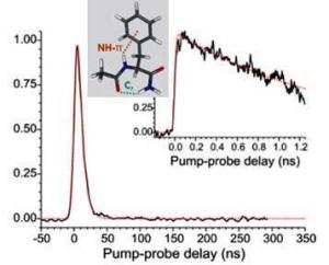
This important issue for biomolecules deals with the relationship between structure (conformation, environment), and dynamics (reactivity, relaxation). This topic is directly connected to the properties of proteins, following electronic excitation consecutive to a near UV photon absorption. Long-lived excited states are indeed potentially harmfull, since they provide opportunity for photochemistry to take place before the molecule relaxes its energy, leading to structural damages potentially affecting the function of the biomolecule. However, it has been suggested from both experimental hints and theoretical evidences that in biomolecules ultrafast relaxation phenomena can take place, allowing them to bypassing damaging photochemical phenomena. This aim of the present work is specifically to document the basic phenomena controlling the lifetime of the excited states, through
- An experimental characterization of the lifetimes, in pump-probe experiments at several timescales (nano-, pico-or femtosecond), and of the nature of the electronic states formed,
- A theoretical modeling of the processes involved, in particular the role of conical intersections among the several electronic states involved.
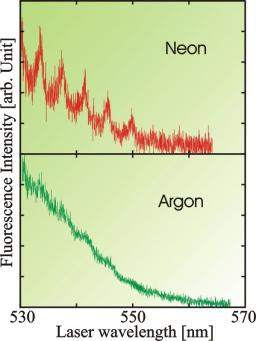
Laser-induced-fluorescence studies of calcium dimer deposited on large argon and neon clusters have been performed. The spectroscopy of the Ca2 A state is slightly perturbed by the cluster surface leading to shifts and broadenings of the order or less than 100 cm-1. An absorption has been evidenced in the 530–550 nm wavelength range that is tentatively assigned to the yet undocumented A’ 1Pu state of Ca2 correlating to the Ca(1D)+Ca(1S) asymptotic limit. The excited calcium dimer dynamics are very different in neon and argon clusters. The argon cluster is much more efficient for electronic and vibrational relaxation of the excited dimer. Finally, excitation in the blue of the calcium atomic resonance line leads to a competition between dissociation of the dimer with ejection of an excited calcium atom out of the cluster and the relaxation of the dimer to lower excited levels.
Spectroscopy and dynamics of calcium dimers deposited on large argon and neon van der Waals clusters
M. A. Gaveau, M. Briant, P. R. Fournier, J. M. Mestdagh, and J. P. Visticot
The Journal of Chemical Physics, 116(3) (2002) 955
|
The generation of radical species triggered by nuclear reactions which produce elevated Linear Energy Transfer (LET) ionizing particles in aqueous media, as well as their reactivity, may be drastically affected by extreme thermodynamic conditions (temperatures higher than 300°C, pressures higher than 100 MPa, acidic or alkaline pH). Such effects are studied by experimental and computational techniques. |
A related project under development concerns the use of new energetic particle sources driven by high intensity lasers. These sources deliver femtosecond bunches of MeV-electrons and also high LET protons, carbons which rendering possible the study of their interaction with various molecular systems, probed by optical spectroscopy. |
Transitions de phase: Une des questions centrales en physique de la matière condensée concerne les transitions de phase entre les états métalliques, isolants et supraconducteurs, dans les matériaux où les corrélations électroniques sont prédominantes. Ces transitions de phase sont accompagnées par des brisures de symétrie structurale et/ou magnétique qui renseignent sur les mécanismes microscopiques à l’origine des corrélations électroniques et des propriétés remarquables de ces composés. Le DRECAM développe des techniques de pointe pour mettre en évidence et caractériser finement les transitions de phase des matériaux.
Conditions extrêmes :Les conditions extrêmes (champs magnétiques, pression, basses températures ou haute densité d’énergie) permettent l’émergence d’effets nouveaux et intéressants dont l’étude renseigne sur les propriétés de la matière dans les grandes profondeurs ou au cœur des planètes géantes gazeuses. Des composés aussi simples et abondants que l’hydrogène et l’oxygène développent à haute pression et à très basse température des phases magnétiques, métalliques ou même supraconductrices inhabituelles. Dans l’oxygène solide sous pression, la structure est stabilisée par les couplages magnétiques dont la force est comparable aux faibles interactions intermoléculaires.
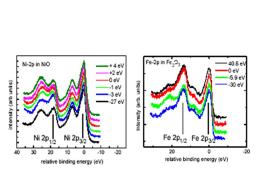
K-edge X-ray absorption and 2p-XPS spectra of 3d-element oxides present spectral features which cannot be explained within a simple one-electron model. These features reveal the fine electronic structure of transition metal (TM) oxides valence states resulting from hybridized TM-3d and O-2p states, and the correlations between these valence electrons. Resonant electronic spectroscopy (resonant Auger or resonant photoelectron spectroscopy) around the TM K-edge can be used to interpret the structures of the threshold and, with the help of theoretical calculations, to determine the electronic configuration of the excited ion. For example, 1s->3d quadrupolar transitions are detected and discriminated from dipolar contributions in the titanium K-edge absorption pre-peaks in TiO2 [1] and in the Ni K-edge prepeaks in NiO thanks to resonant KLL Auger spectra. [2] The experimental results obtained on Ti K-edge are successfully reproduced by theoretical calculations [3], making the determination of some electronic parameters possible from experimental data.
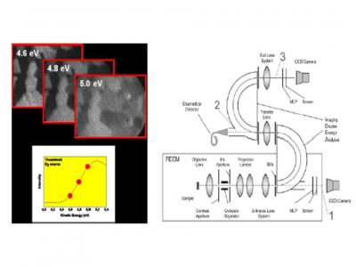
Information for the assignment of fine structure in the absorption can be also deduced from hard X-ray resonant XPS. This has been demonstrated at the L3 edge of intermediate valence Ce compounds where the contributions of the 4f0, 4f1 and 4f2 electronic configurations have been located by resonant Ce-3d photoelectron spectroscopy. [3] In 3d-TM oxides, the 2p-XPS spectra also contain valuable information. The main line can be split into two components (like in NiO) revealing non-local core hole screening effects and satellites appear due to the different valence configurations. Can these spectral features be used to explore the electronic configurations of the different final states reached through the absorption edge? To explore this possibility, we have studied the resonance of 2p-XPS around the TM K-edge in NiO and Fe2O3 (Fig. 1). [2] One observes the same behaviour on the two samples: the relative intensity of the 2p1/2 main peak resonates at a photon energy below the white line (maximum of the absorption) (at -3 eV for NiO and -5 eV for Fe2O3), and the 2p1/2 satellite is maximum for a photon energy slightly higher than the white line. The resonant effects are more pronounced on NiO than on Fe2O3, and no resonant effects are observed on TiO2 around the Ti K edge. This can be related to higher localisation of valence electron as the number of 3d electrons is increased, and a more “atomic-like” behaviour.
REFERENCES :
[1] J. Danger et al., Phys.Rev Lett. 88, 243001 (2002)
[2] P. Le Fèvre et al, . Nucl. Instr Meth A 547, 176 (2005)
[3] P. Le Fèvre et al., J. Electron Spectrosc. Relat. Phenom. 136, 37 (2004)
The NANOAM project (Nanometer scale induced nanostructure between amorphous layers and crystalline materials) was a collaboration between five european laboratories, funded by the 5th PCRD, and six laboratories from the US, funded by the NSF. The work program was mainly focused on silicate materials, and we were mainly involved in two workpackages, centered on :
1 - the interface between thin silicate amorphous films and a silicon single crystalline substrate, with expected applications in the field of high-κ dielectrics, possibly able to replace ultra-thin silicon oxide films in future electronic devices ;
2 - the nanoscale amorphous films which form at the grain boundaries of silicon nitride-based ceramics, when rare earth (RE) oxides are introduced as sintering aids. As these films have a strong influence on the resulting properties of the ceramic, the ultimate goal was grain boundary engineering, in order to optimize the properties of the ceramic by controlling the nanostructure of these films.
The workpackages were equally balanced between theoretical approaches (electronic structure calculations, phase field modelling, molecular dynamics…) and experimental determination of the structure and dielectric properties of these amorphous films, by using mainly the recent developments of transmission electron microscopy and related techniques such as transmission electron energy loss spectroscopy (TEELS). Our contribution to this collaborative work was to perform X-ray Photoelectron Spectroscopy (XPS) and Reflection Electron Energy Loss Spectroscopy (REELS) on nanometer-scale amorphous silicon oxide films (WP1) [1,2], and on reference materials in the bulk form, such as oxynitride glasses, with the same structure and composition as the one of intergranular glassy films (WP2).
Silicon oxide films
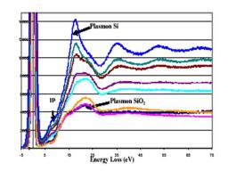
A central goal of the research undertaken was to determine whether Si-O films on silicon could exhibit equilibrium self-selecting thickness and composition that may be systematically controlled by the oxygen activity in the system. A new approach to growing stable Si-O films on silicon was therefore being explored at MIT using a metal/metal oxide (Zr/ZrO2 or TiO/Ti2O3) buffer system to control the oxygen activity (aO2) at extremely low levels. An iterative series of synthesis and characterization experiments between our group and MIT was conducted. The thickness of the films was measured from the intensity ratio from the pure Si and Si-O components of the Si2p photoelectron line, as well as from REELS experiments at different energies (Fig. 1). Experimental results in the synthesis and characterization of films provided compelling support for the existence of stable nanoscale films on silicon, with a thickness in the nm range, corresponding to an equilibrium under the given annealing conditions for each oxygen activity. This potentially offers a new approach to controlled fabrication of interfacial oxides in silicon microelectronics (gate oxides).
Oxynitride glasses
Two series of oxynitride glasses elaborated respectively at Karlsruhe University (RE-Si-Mg-O-N) and at Oak Ridge National Laboratory (RE-Si-Al-O-N) have been studied by XPS and REELS. The oxygen 1s photoelectron line-shape reveals a striking difference between the La and Lu glasses: the O1s photoelectron lines of the La glasses are broader than the ones of the Lu glasses (Fig. 2). This broadening comes from an increased contribution of O-Si bonds compared to the Lu glasses. This result is an experimental evidence that Lu has a larger affinity for oxygen versus nitrogen than La, as theoretically predicted by ab initio calculations. [3] La has an increased preference for N environment, compared to Lu, so that more O-Si bonds are expected in the La containing glasses than in the Lu containing glasses.
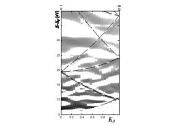
In photoemission, the initial-state k⊥ can be deduced if the final-state surface-perpendicular dispersion E(k⊥) into the crystal bulk is known. The widely used free-electron-like (FE-like) model fails for many quasi 2D systems.[1] Final states can feature dramatic non-free-electron effects, [2] and can undergo significant energy shifts due to band- and k-dependent excited-state self-energy corrections ΔΣ.[3] The photoelectron lifetime damps the final-state wavefunction in the surface-perpendicular direction towards the crystal interior, resulting in intrinsic final-state broadening in k⊥.[4] The PE peaks then reflect the initial-state E(k⊥) average over the broadening interval.
True final-state E(k⊥) dispersions can be obtained using Very-Low-Energy Electron Diffraction (VLEED). In the one-step PE theory the final state can be considered as the time-reversed LEED state. In VLEED the energies of the spectral structures reflect the characteristic points in the final-state E(k⊥) such as the band gap edges, and their broadening and relative amplitudes the corresponding lifetimes. [5]
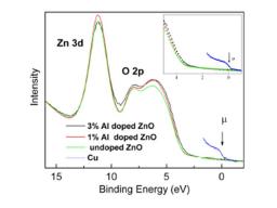
Transparent conductive oxide (TCO) films have been extensively used in optoelectronic devices because of their high visible transmittance and low DC resistivity. ZnO has appropriate optical transparency and small reflectance at 600 nm, the optimal wavelength for the silicon solar cells [1], can be used as the passivation layer of the active Si electrode and is naturally abundant and non toxic. The use of a single layer acting simultaneously as the antireflection coating, the passivation layer and the electrode for extracting the electrons from the photovoltaic cell, could be a real breakthrough in the solar cell technology, simplifying fabrication and reducing costs. Spray pyrolysis is an interesting alternative method to obtain TCO coatings because of its simplicity: it can coat extended surfaces; does not need high vacuum, and is easily inserted in a production line. [2]
Increases in conductivity of several orders of magnitude have been observed when doping ZnO films with a group III element up to a [Al]/[Zn] = 3% atomic ratio. An open question is the conduction mechanism compared to undoped films.
Resistivity of the films was determined using Van der Pauw’s method. A decrease of resistivity in two orders of magnitude is measured for 3% Al doping. The high resolution valence-band PES spectra using 35eV photons are shown in Fig. 1 (right). [3] None of the samples present a density of states in the band gap region. We observe a slight reduction of the band gap compared to the 1% and the undoped samples. The band gap is reduced by ~150 meV. This is interpreted as a result of the hybridization between the Al-dopants with the O atoms in the ZnO. A slight decrease of the optical band-gap (~ 90 meV) is also measured in transmittance measurements. [4] No insulator-metal transition is observed. Conductivity occurs principally via a band conduction mechanism. The conductivity jump is described in terms of a Vervey transition.[5] The independent variable is the Al doping, giving discontinuity in the first derivative of Gibb’s function Δσ/ΔND. ΔVBM = 150 meV
leads to a decrease ΔEA in the activation energy for electrons to accede to the conduction band. Thus the conductivity jump Δσ can be written as Δσ ≈ exp (150/kT) = 378, similar to the electrical measurements.
REFERENCES :
[1] J.J. Aranovich et al., J. Appl.Phys. 51, 4260-4268 (1980)
[2] H. Mondragón-Suárez et al., Appl. Surf. Sci. 193, 52-59 (2002)
[3] M. Addou et al., J. Chim. Phys. Phys.Chim. Biol. 96, 232-244 (1999)
[4] J.H. Park et al., Phys. Rev. B 55, 12813-12817 (1997)
MEcanismes de la Solubilisation ENzymatique des GEls
Construction d'un rhéomètre parallèle
L'invasion cellulaire est le processus par lequel des cellules malignes quittent un cancer primaire pour passer dans la circulation sanguine et former des métastases à distance de la tumeur d'origine. La formation de ces métastases est bien sûr un facteur essentiel du pronostic. Une des étapes de ce phénomène est le passage des cellules à travers la matrice extra cellulaire. Cette matrice a pour base mécanique un gel de collagène dont la cellule invasive doit couper les chaînes pour se frayer un passage. Nous avons entrepris d'étudier les aspects de ce processus relevant de la physique de la « matière molle » et le présent projet s'inscrit dans ce contexte.
Une des difficultés principales rencontrées lors d'une étude expérimentale « physique » de systèmes biologiques est la variabilité intrinsèque de ces systèmes qui impose, pour obtenir une image pertinente du phénomène, une multiplication des échantillons mesurés et donc un dispositif expérimental très simple pouvant être multiplié également. L'étude [1] que nous avons publiée sur ce sujet reposait sur ce principe : Le dispositif expérimental unitaire était constitué d'un récipient ( tube de centrifugation ) contenant le gel et l'enzyme dissous et une bille de plastique. Un grand nombre de ces tubes était placé dans un thermostat et observé par un unique appareil photo numérique. La donnée relevée était la durée totale de la dégradation révélée par la chute de la bille.
Le résultat le plus déconcertant de cette étude était que ce temps de dégradation ne varie pas comme une puissance entière de la concentration en enzyme, mais comme une puissance -3/2 environ. L'intuition et tous les modèles simples prévoient une proportionnalité inverse. Le seul modèle (à notre connaissance) qui permette d'expliquer cet exposant est le suivant : chaque enzyme reste confinée dans une « bulle » sphérique qu'elle crée et ces bulles percolent (percolation continue ou « fromage suisse »). Afin que la bulle reste sphérique, l'enzyme doit suivre une marche auto-attirante où la probabilité de pénétrer en territoire vierge est inférieure à un certain seuil. Dans ce modèle, l'exposant asymptotique est de 4/3, mais il est atteint par au-dessus et passe par 3/2 pour certaines valeurs finies.
Un phénomène moléculaire a pu être établi (ou fortement suggéré) par des mesures macroscopiques interprétées par des modèles de la physique statistique.
Quelques mesures de constantes élastiques de gels en cours de dégradation ont été effectuées et montrent des variations en loi de puissance, en accord, au moins qualitativement, avec les résultats précédents. Ces mesures se font avec un instrument relativement complexe qu'il est donc impossible de multiplier.
Des expériences préliminaires mettant en jeu d'autres couples enzyme-gel ont montré des exposants différents. Les problèmes tant biologiques que physiques sont donc plus généraux que celui considéré jusqu'ici. Des moyens expérimentaux et théoriques supplémentaires seraient utiles.
Le premier objectif du présent projet est de construire un montage expérimental permettant une mesure de l'élasticité du gel suffisamment simple pour être répliqué en plusieurs exemplaires.
Présentation PowerPoint
The present work is at the interface of studies of fracture and structure of materials. What can be known from a fracture surface? Can we improve our knowledge of a material structure from its fracture surfaces? Fracture surfaces of a quasicrystal are here considered.
On the quasicrystal side (QC), aperiodic materials can be seen as the projection in three dimensions of a periodic structure in a virtual space of higher dimension (6 for QC with icosahedral (i-) symmetry). In real space, the structure may be described as an aperiodic assembly of overlapping, so called Bergman or Mackay, clusters. This very cluster structure was suggested to be at the origin of the mysterious stability of QCs. Clusters being able to be preserved in a fracture experiment, analysis of fracture surfaces are an attempt to found evidence of their relevance in the structure.
On the fracture side, roughness of the fracture surface of various heterogeneous materials (Al alloy, mortar, silica glass…) was shown to be self-similar whatever the heterogeneity characteristic length is. This universal behavior with z = 0.8 power law dependence is pointed out from the elementary grain scale up to the process zone size given by  . Accordingly, i-AlPdMn QC offers unexplored conditions: the extension of the process zone (estimated from the toughness ΚIc and the upper yield stress σY at room T) is of the order of ~ 0.09 nm, lower than the atomic size, making i-AlPdMn a hyper-brittle material.
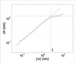

Introduction
Many industrial applications would benefit greatly of further insight into the mixing mechanisms of viscous fluids. The study of fluid mixing enhanced by closed flows goes back to the early 80’ s and the introduction of chaotic advection. The interest for mixing in open flows is more recent. Transient mixing created by an open flow whose time dependency is restricted to a bounded region – the mixing region – can be studied in the same dynamical systems framework as for bounded flows, since for typical open flows, Lagrangian trajectories have a transient chaotic behavior inside the mixing region. Nevertheless, quantitative analysis of homogenization realized by such flows is still lacking. We have characterized transient mixing in open flows and in parallel, we carried complementary investigations in closed flows as a guide on the less-beaten track of open flows.
Our study, experimental, numerical and theoretical, is based on two types of chaotic mixing experiments, in a closed vat and in an open channel. For closed flows, we have first proposed a topological description of mixing by the entanglement of periodic orbits that we called ”ghost rods”. The experimental study of the concentration field of a dye has then revealed the role of the walls of the domain where mixing takes place, in closed and open flows as well. For closed flows, the chaotic or regular nature of trajectories initialized close to the wall determines the evolution of the concentration field, even far from the walls. In particular, we have observed slow (algebraic) dynamics of homogenization when the chaotic region extends to no-slip walls. In open flows, we have reported on the evolution of the concentration field in the mixing and downstream regions, resulting from the injection of a dye blob. We have described the poorly mixed elements that escape quickly, as well as the asymptotic onset of a permanent (self-similar) pattern determined by the periodic orbits inside the mixing region. Finally, various models derived from the baker’s map have allowed us to understand most observed mechanisms.
A number of experimental works have studied surface instability of a granular heap, namely avalanches, and have led to the conclusion that such avalanches do not behave in a critical manner at all. In recent years, it has also been suggested that the jamming transition of granular materials could be analog to a glass transition. Further works on mean field glass models have developed this analogy and tried to unify concepts that had emerged in both fields, such as the dynamical temperature defined from fluctuation dissipation relations, and that related to Edwards' statistical ensemble.
First, the micro-displacements generated by a small localized overload at the free surface are visualized experimentally inside a packing of steel beads. For a triangular packing, beads rearrangements remain confined in two inverted triangles on both sides of the applied overload. This pattern disappears for stronger disorder. A simple model allows us to account for these observations and to relate them to the stress function response measured via photoelastic visualizations.

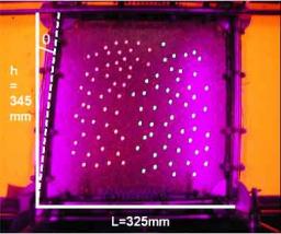
Granular media involve many particles, so that it is tempting to describe them thermodynamically. Unfortunately, energy is lost through friction and has to be brought by a non-thermal source. As a consequence, the dynamical equations do not leave any obvious ensemble invariant.
However it has been proposed that these systems could have a statistical mechanics of their own. Here, we present experimental results on the statistical properties of 2D disordered granular media under cyclic shear in an attempt to check the possibility of a thermodynamic construction for dense granular materials.
Dunes dynamics
Dunes dynamics has strong impact on the ecology and the economy of sandy areas, but is far from being understood. Since the work on sand dune formation by R.A. Bagnold, a world wide inventory of dunes has been developed. The most common dune, the so-called barchan, has a typical crescent shape normal to the wind, with arms downwind and forms under mono-directional winds. Various models call for more experimental data. On one hand, field measurements are difficult to perform and often incomplete. On the other hand, it is believed that dunes have a minimal size of the order of one meter, not reducible to smaller laboratory scales. We have set up a wind channel where to conduct experiments with sand and we have shown experimentally that an initial sand pile, under a wind flow charged in sand, flattens and exhibits a typical barchan shape before disappearing. An evolution law has been proposed for the profile and the summit of the dune. The dune dynamics is shape invariant. The invariant shape, the "dune function", was isolated. Our results, which clearly demonstrates the feasibility of dunes in the lab, open a new way of investigations in desert studies.
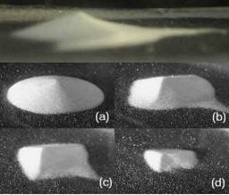
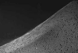
Surface flows
We investigate steady granular surface flows in a rotating drum, measuring the velocity profile in the flowing layer at the center of the drum, the flowing layer thickness and the static/flowing boundary profiles. The velocity varies linearly with depth, with a gradient independent of both the flowing layer thickness and the static/flowing boundary local slope. Then, writing the depth averaged conservation equations for granular surface flows, it was possible to find experimentally the constitutive relations needed to close these equations. Altogether it has brought the evidence that the relation between stress and strain is non local:
In photoemission, the initial-state k⊥ can be deduced if the final-state surface-perpendicular dispersion E(k⊥) into the crystal bulk is known. The widely used free-electron-like (FE-like) model fails for many quasi 2D systems.[1] Final states can feature dramatic non-free-electron effects, [2] and can undergo significant energy shifts due to band- and k-dependent excited-state self-energy corrections ΔΣ.[3] The photoelectron lifetime damps the final-state wavefunction in the surface-perpendicular direction towards the crystal interior, resulting in intrinsic final-state broadening in k⊥.[4] The PE peaks then reflect the initial-state E(k⊥) average over the broadening interval.
True final-state E(k⊥) dispersions can be obtained using Very-Low-Energy Electron Diffraction (VLEED). In the one-step PE theory the final state can be considered as the time-reversed LEED state. In VLEED the energies of the spectral structures reflect the characteristic points in the final-state E(k⊥) such as the band gap edges, and their broadening and relative amplitudes the corresponding lifetimes. [5]
The NANOAM project (Nanometer scale induced nanostructure between amorphous layers and crystalline materials) was a collaboration between five european laboratories, funded by the 5th PCRD, and six laboratories from the US, funded by the NSF. The work program was mainly focused on silicate materials, and we were mainly involved in two workpackages, centered on :
1 - the interface between thin silicate amorphous films and a silicon single crystalline substrate, with expected applications in the field of high-κ dielectrics, possibly able to replace ultra-thin silicon oxide films in future electronic devices ;
2 - the nanoscale amorphous films which form at the grain boundaries of silicon nitride-based ceramics, when rare earth (RE) oxides are introduced as sintering aids. As these films have a strong influence on the resulting properties of the ceramic, the ultimate goal was grain boundary engineering, in order to optimize the properties of the ceramic by controlling the nanostructure of these films.
The workpackages were equally balanced between theoretical approaches (electronic structure calculations, phase field modelling, molecular dynamics…) and experimental determination of the structure and dielectric properties of these amorphous films, by using mainly the recent developments of transmission electron microscopy and related techniques such as transmission electron energy loss spectroscopy (TEELS). Our contribution to this collaborative work was to perform X-ray Photoelectron Spectroscopy (XPS) and Reflection Electron Energy Loss Spectroscopy (REELS) on nanometer-scale amorphous silicon oxide films (WP1) [1,2], and on reference materials in the bulk form, such as oxynitride glasses, with the same structure and composition as the one of intergranular glassy films (WP2).
Silicon oxide films
A central goal of the research undertaken was to determine whether Si-O films on silicon could exhibit equilibrium self-selecting thickness and composition that may be systematically controlled by the oxygen activity in the system. A new approach to growing stable Si-O films on silicon was therefore being explored at MIT using a metal/metal oxide (Zr/ZrO2 or TiO/Ti2O3) buffer system to control the oxygen activity (aO2) at extremely low levels. An iterative series of synthesis and characterization experiments between our group and MIT was conducted. The thickness of the films was measured from the intensity ratio from the pure Si and Si-O components of the Si2p photoelectron line, as well as from REELS experiments at different energies (Fig. 1). Experimental results in the synthesis and characterization of films provided compelling support for the existence of stable nanoscale films on silicon, with a thickness in the nm range, corresponding to an equilibrium under the given annealing conditions for each oxygen activity. This potentially offers a new approach to controlled fabrication of interfacial oxides in silicon microelectronics (gate oxides).
Oxynitride glasses
Two series of oxynitride glasses elaborated respectively at Karlsruhe University (RE-Si-Mg-O-N) and at Oak Ridge National Laboratory (RE-Si-Al-O-N) have been studied by XPS and REELS. The oxygen 1s photoelectron line-shape reveals a striking difference between the La and Lu glasses: the O1s photoelectron lines of the La glasses are broader than the ones of the Lu glasses (Fig. 2). This broadening comes from an increased contribution of O-Si bonds compared to the Lu glasses. This result is an experimental evidence that Lu has a larger affinity for oxygen versus nitrogen than La, as theoretically predicted by ab initio calculations. [3] La has an increased preference for N environment, compared to Lu, so that more O-Si bonds are expected in the La containing glasses than in the Lu containing glasses.
K-edge X-ray absorption and 2p-XPS spectra of 3d-element oxides present spectral features which cannot be explained within a simple one-electron model. These features reveal the fine electronic structure of transition metal (TM) oxides valence states resulting from hybridized TM-3d and O-2p states, and the correlations between these valence electrons. Resonant electronic spectroscopy (resonant Auger or resonant photoelectron spectroscopy) around the TM K-edge can be used to interpret the structures of the threshold and, with the help of theoretical calculations, to determine the electronic configuration of the excited ion. For example, 1s->3d quadrupolar transitions are detected and discriminated from dipolar contributions in the titanium K-edge absorption pre-peaks in TiO2 [1] and in the Ni K-edge prepeaks in NiO thanks to resonant KLL Auger spectra. [2] The experimental results obtained on Ti K-edge are successfully reproduced by theoretical calculations [3], making the determination of some electronic parameters possible from experimental data.
Information for the assignment of fine structure in the absorption can be also deduced from hard X-ray resonant XPS. This has been demonstrated at the L3 edge of intermediate valence Ce compounds where the contributions of the 4f0, 4f1 and 4f2 electronic configurations have been located by resonant Ce-3d photoelectron spectroscopy. [3] In 3d-TM oxides, the 2p-XPS spectra also contain valuable information. The main line can be split into two components (like in NiO) revealing non-local core hole screening effects and satellites appear due to the different valence configurations. Can these spectral features be used to explore the electronic configurations of the different final states reached through the absorption edge? To explore this possibility, we have studied the resonance of 2p-XPS around the TM K-edge in NiO and Fe2O3 (Fig. 1). [2] One observes the same behaviour on the two samples: the relative intensity of the 2p1/2 main peak resonates at a photon energy below the white line (maximum of the absorption) (at -3 eV for NiO and -5 eV for Fe2O3), and the 2p1/2 satellite is maximum for a photon energy slightly higher than the white line. The resonant effects are more pronounced on NiO than on Fe2O3, and no resonant effects are observed on TiO2 around the Ti K edge. This can be related to higher localisation of valence electron as the number of 3d electrons is increased, and a more “atomic-like” behaviour.
REFERENCES :
[1] J. Danger et al., Phys.Rev Lett. 88, 243001 (2002)
[2] P. Le Fèvre et al, . Nucl. Instr Meth A 547, 176 (2005)
[3] P. Le Fèvre et al., J. Electron Spectrosc. Relat. Phenom. 136, 37 (2004)
During the preparation of ultra flat surfaces of stainless steel for the two above studies of degradation of these materials, we experimented an electrochemical polishing process with a classical bath for obtaining a mirror-like surface. The electrochemical solution consists in perchloric acid in an organic solvent cooled at constant temperature ~ 4°C. Either the current ranging from 0.1 to 0.8A or the voltage varying from 15 to 60V was controlled in the electrochemical cell. AFM images of the electropolished surface showed an unexpected nanostructure. [1] As shown in Fig. 1 (a), the surface is covered by a quasi periodic arrangement of pores with a typical diameter of 80nm and a period of 100-120nm. Large scan images reveal that the arrangement of pores forms grains with grain boundaries and defects as dislocations. Interestingly, this lattice is independent of the grain orientation of the underlying metal. The chemical nature of this overlayer analyzed by X-ray photoemission brings
evidence that the surface layer is formed by chromium and iron oxides. No signal coming from nickel atoms was detected. Changing the electrochemical conditions (temperature, current or voltage) influences the pore diameter and the arrangement period which varies from 50 to 150 nm.
This nanostructured surface appears well suited for improving the quality of osteointegration of stainless steel prosthesis in bones since rough-surfaced implants favour both bone anchoring and biomechanical stability. In collaboration with an INSERM group (Nantes), which studies the interface between bone and metal implants, the anchoring of osteoblast cells onto these nanostructured surfaces of stainless steel was investigated in vitro. The first results shown in Fig.1 (b) have demonstrated that the quality of the cell attachment on these nanostructured surfaces is better than on rough surfaces of stainless steels prepared with standard processes (mechanical abrasion, sanded surfaces,…). [1]
REFERENCES :
[1] F. A. Martin, Ph.D Thesis Paris VI (2005)
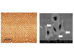
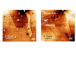
In this work, damages induced by stress were investigated in situ by using the set up described above, which combines an AFM microscope working in contact mode, an electrochemical cell and a tensile micromachine. First, the changes in the surface morphology induced by plastic deformation of the specimen will be described and analysed. Then, we will report the first observation of nanometre damages generated by a stress in a corrosive environment.
As in the corrosion study presented above, two kinds of stainless steels were studied: austenitic steel (304L) and a duplex one containing an austenite and a ferrite phases (Uranus 50). The preparation of specimen presents many stages including mechanical and electrochemical polishing with a final mechano-chemical polishing. From AFM micrographs 5μm x 5μm in size, the best results characterizing the surface roughness are ~ RMS 0.5nm but the surface of some specimens present ripples.
It is well known that plastic deformation generates dislocations in crystals whose some of them emerge at the surface. The evolution of the surface morphologies of the two kinds of stainless steels has been
explored by in situ AFM during the plastic deformation. After the tensile test, Electron Back Scattered Diffraction (EBSD) measurements were performed on the area observed by AFM.
Fig. 1 presents examples of AFM micrographs documenting the evolution of the surface relief topography during the tensile test of the duplex steel. Before the tensile test, the different grains can be easily recognized from the orientation of ripples for the austenite grains. The ferrite grains, which are harder than the austenite ones appear clearer in the AFM images. This means that the preparation of the specimen surface yields prominent ferrite grains. After 0.6mm elongation of the sample, some austenite grains exhibit nearly equidistant lines, which correspond to the intersection of active slip planes with the grain surface (Fig. 1b). Indeed, these lines due to the emergence of dislocations are steps whose height can be accurately measured. After 0.8 mm elongation, both the density and theheight of the steps increase when compared to those observed in the 0.6mm elongation image. We also pointed out that a secondary slip system is activated in some austenite grains. In this image, no sign of plastic deformation was detected in the ferrite grains. Elsewhere on the surface, we observed the presence of steps in ferrite grains. These steps appear connected to the accumulation of dislocations in a neighbouring austenite grain.
The early stages of corrosion of stainless steels under stress are still not very well understood despite a lot of investigations. [1] Many techniques have been used for studying this process of metal damaging because of its huge economical importance. However, none of these investigations, except optical microscopy, implies in situ observations and provides maps of damaged areas. Thus, the degradation if these metals was studied with a poor resolution, typically above the micrometre. As a consequence, the onset of corrosion is believed to appear randomly along the metal surface. The uprising of local probe microscopy in the 90’s and specially the atomic force microscopy (AFM) has opened new avenues for in situ exploration of these damaging processes at sub micrometre scale.
In this work, damages induced by corrosion were investigated in situ by using a new and original setup, which combines an AFM microscope working in contact mode and an electrochemical cell. We will present surface modifications due to corrosion without an external stress applied to the sample.
Two kinds of stainless steels were studied: an austenitic steel (304L) and a duplex one containing an austenite and a ferrite phases (Uranus 50). As we want to detect defects in the nanometre range, the specimen preparation presents many stages including mechanical and electrochemical polishing with a final mechano-chemical polishing. This last treatment has been performed with colloidal silica particles for about 20 h. From AFM micrographs 5μm x 5μm in size, the best results characterizing the surface roughness are ~ RMS 0.5nm.
The first stages of corrosion of stainless steels covered by a passive layer have been studied by in situ AFM in chloride containing solutions at room temperature. The solution contains 0.5M of NaCl and 0.01M of borate salts. The specimen was immersed in solution and a - 400 mV potential (vs.Ag/AgCl) was applied during 300s. Then the potential was raised at a low rate (1mV s-1) until pitting occurred as shown by the intensity increase in the electrochemical cell. Both 304L and Uranus 50 stainless steels exhibit a free corrosion potential at -230 ± 25mV (vs. Ag/AgCl) in these operating conditions.
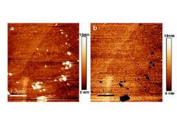
Photoelectron emission microscopy combined with intense photon sources is becoming a powerful analytical tool with both spatial resolution down to a few nanometers and spectroscopic capabilities. One can laterally resolve surface domains according to their magnetization, chemical composition, electronic structure, and even follow the dynamics of domain movement, catalysis or laser emissions using time resolved detection systems.
The lateral resolution varies from 1 mm to 10 nm, placing this technique at the heart of the mesoscopic physics, and corresponds to the need for spectroscopic information on the intermediate scale between atomic and macroscopic, so lacking in multiscale modeling. Furthermore, the possibility of studying nanostructured materials and objects on the 100 nm scale corresponds perfectly to the technologically interesting length scale in electronics, bioelectronics.
CEA has acquired the first production model of the NanoESCA (OMICRON GmbH), a spectromicroscope designed for high spatial and energy resolution over a wide range of photoelectron kinetic energies (Δx ~ 100 nm; ΔE ~ 100 meV). This is done thanks to the combination of a simple purely electrostatic lens column allowing accurate focus tracking over several hundred eV with the use of a double hemispherical analyzer, eliminating spherical aberrations usually present in energy analyzers for photoelectron microscopes.
This research is carried within the framework of the 6th PCRD INCEMS project. [1] The objective is to predict the properties of multifunctional ceramics in the presence of Intergranular films (IGFs). Regrouping expert theoretical and experimental groups from Germany, UK, Slovenia and France, the CEA contributes the possibility of spectroscopic imaging on the mesoscopic scale, providing essential experimental verification of the extension of atomistic models to larger scales.
The electrical properties of electro-ceramics are largely governed by their complex microstructure and in particular by the grain boundaries, and must be precisely controlled for their applications (as, for instance, sensors, actuators and capacitors). A recent study has furnished first evidence for IGFs in SrTiO3.[2]
In this study, we have studied the dependence on niobium segregation in strontium titanate of various sintering parameters. 1.2 mol% Nb doped SrTiO3 was sintered at 1420°C in air for different times and with different post sintering cooling rates. Scanning electron microscopy images provided evidence of morphological differences among the prepared samples. We have established a surface preparation protocol from a combined XPS/AES/LEED study to obtain stoichiometric surfaces, important for reliable analysis of the electrical properties such as the work function. In particular we have proven that the often used oxygen plasma is not suitable.
An accurate X-ray photoemission study has shown that for 20 hours sintering in a reducing atmosphere there is the appearance of TiOx localized near the surface, either as a grain termination layer or in intergranular regions, as suggested by SEM.
Thirdly we correlate a core level shift (ΔE = 0.24 eV) in the Nb 5d3/2 with short sintering times. The smaller grain size, allows higher Nb segregation to the grain boundary region, corroborated by the decreased lattice parameter measured by X-ray diffraction due to niobium leaving titanium substitutional sites and going to grain boundary zones. We have obtained the first laterally resolved photoemission threshold images in XPEEM using the NanoESCA machine in laboratory conditions (see §1.10 below), suggesting up to four different work functions (Δ ΦWF = 0.37 eV). Electron back-scattering and synchrotron radiation induced XPEEM are in progress to verify this.
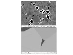

Silicon nanocrystals (Si NCs) memories are currently studied as solutions to overcome the scaling limitations of conventional flash and non-volatile memories.[1,2] High density Si NCs (~ 5nm diameter) on a low leakage tunnelling dielectric give a memory device with a large threshold voltage shift and long time retention. [3,4]
Si NCs were grown by LPCVD on 3 nm-thick Al2O3 layers deposited onto oxidized Si by Atomic Layer deposition.[5] The growth conditions for the Si NCs yielded surface coverage of around 30 % and an average diameter of 5 nm. Synchrotron radiation X-ray photoelectron spectroscopy (SRXPS) is used for the study of Si nano-crystals (NCs) for applications in non-volatile memory devices. The tunability of the photon energy allows selective characterization either of the oxidation shell or of the core of the NCs.
No exact analytical solution exists for hemispherical dots based on the approach of Himpsel et al.[6] We have used a rectangular profile as an approximation to the hemisphere, allowing an analytical integration over x and z, with a correction factor in order to extract the oxide shell thickness. [7]
We observe chemical shifts for the oxidized components that are consistent with those observed for planar oxidation. The FWHMs are much larger for the NCs than for a planar surface. This broadening is clear on the spectra for the Si4+ peak, and confirmed for every spectral component, including Si0; we observe for the NCs Si 2p components a Gaussian width which is 0.35-0.40 eV larger than for the planar oxidized Si.
Inhomogeneous stress in the NC volume upon oxidation gives structural disorder. [8] The core and the oxide shell present different stress state, and even in the shell itself the stress may depend on the thickness.
REFERENCES :
[1] M. L. Ostraat et al,. Appl. Phys. Lett. 79, 433 (2001)
[2] G. Ammendola et al., Solid-State Electron. 48, 1483 (2004)
[3] T. Baron et al. , Appl. Phys. Lett. 82, 4151 (2003)
[4] S. Tiwari et al,. Appl. Phys. Lett. 68, 1377 (1996)
[5] O. Renault et al,. J. Vac. Sci. Technol. A 20, 1867 (2002)
[6] J. Himpsel et al,. Phys. Rev. B 38, 6084 (1988)
[7] O. Renault et al, Surf. Inter. Anal. 38, 486 (2006)
[8] J. Dalla Torre et al,. J. Appl. Phys. 92, 1084 (2002)
High-κ dielectric materials are required to replace SiO2 (κ=3.9) in sub-0.1 μm CMOS, providing better insulating properties: reduced electron tunneling with capacitance characteristics of a much thinner SiO2, thus yielding a very thin equivalent oxide thickness (EOT). [1] Hafnium oxide (HfO2) and its silicates (HfxSi(1-x)O2) have already demonstrated the best compatibility with the main electrical requirements of advanced CMOS technology favouring low leakage currents (conduction band offset ΔEc > 1 eV).
We first investigated and verified the formation of thin interface silicate between a HfO2 high-κ oxideand SiO2 bottom layer, necessary to minimize interface states. We show that the silicate layer may be restricted to two atomic layers, thus playing a negligible role in determining the electronic properties of the stack. Between HfO2 and Si, the valence- and conduction-band offset are determined as 3.2 and 1.4 eV respectively. The thermal stability of the interfacial silicate layer was also addressed. By a quantitative treatment of the Si 2p core-level intensities, we conclude that post deposition annealing under nitrogen leads to the extension of both the interface silicate and the SiO2 interface oxide.
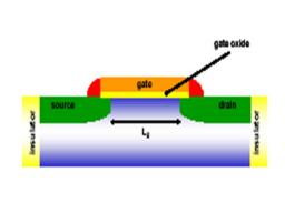
Transparent conductive oxide (TCO) films have been extensively used in optoelectronic devices because of their high visible transmittance and low DC resistivity. ZnO has appropriate optical transparency and small reflectance at 600 nm, the optimal wavelength for the silicon solar cells [1], can be used as the passivation layer of the active Si electrode and is naturally abundant and non toxic. The use of a single layer acting simultaneously as the antireflection coating, the passivation layer and the electrode for extracting the electrons from the photovoltaic cell, could be a real breakthrough in the solar cell technology, simplifying fabrication and reducing costs. Spray pyrolysis is an interesting alternative method to obtain TCO coatings because of its simplicity: it can coat extended surfaces; does not need high vacuum, and is easily inserted in a production line. [2]
Increases in conductivity of several orders of magnitude have been observed when doping ZnO films with a group III element up to a [Al]/[Zn] = 3% atomic ratio. An open question is the conduction mechanism compared to undoped films.
Resistivity of the films was determined using Van der Pauw’s method. A decrease of resistivity in two orders of magnitude is measured for 3% Al doping. The high resolution valence-band PES spectra using 35eV photons are shown in Fig. 1 (right). [3] None of the samples present a density of states in the band gap region. We observe a slight reduction of the band gap compared to the 1% and the undoped samples. The band gap is reduced by ~150 meV. This is interpreted as a result of the hybridization between the Al-dopants with the O atoms in the ZnO. A slight decrease of the optical band-gap (~ 90 meV) is also measured in transmittance measurements. [4] No insulator-metal transition is observed. Conductivity occurs principally via a band conduction mechanism. The conductivity jump is described in terms of a Vervey transition.[5] The independent variable is the Al doping, giving discontinuity in the first derivative of Gibb’s function Δσ/ΔND. ΔVBM = 150 meV
leads to a decrease ΔEA in the activation energy for electrons to accede to the conduction band. Thus the conductivity jump Δσ can be written as Δσ ≈ exp (150/kT) = 378, similar to the electrical measurements.
REFERENCES :
[1] J.J. Aranovich et al., J. Appl.Phys. 51, 4260-4268 (1980)
[2] H. Mondragón-Suárez et al., Appl. Surf. Sci. 193, 52-59 (2002)
[3] M. Addou et al., J. Chim. Phys. Phys.Chim. Biol. 96, 232-244 (1999)
[4] J.H. Park et al., Phys. Rev. B 55, 12813-12817 (1997)
