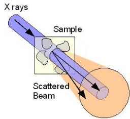
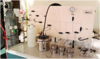
X-rays are used to investigate the structural properties of solids, liquids or gels. Photons interact with electrons, and provide information about the fluctuations of electronic densities in heterogeneous matter. A typical experimental set-up is shown on Figure : a monochromatic beam of incident wave vector is selected and falls on the sample. The scattered intensity is collected as a function of the so-called scattering angle 2 teta.
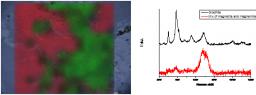
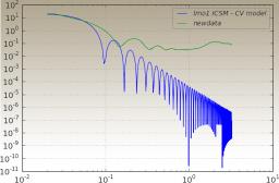
A high sensitivity Small Angle X-Ray Scattering (SAXS) experiment
High resolution calibrated Ultra Small Angle X-ray Scattering (USAXS) on a laboratory source
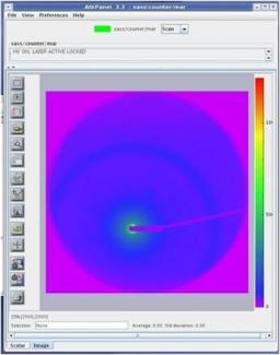
Laboratory experiments are increasingly composed of electronic hardware and computer controlled. In most cases the quality of the equipment (mechanical, electronics) is very good but the software is inadequate and not very flexible to the needs of researchers.
LIONS chose to use the open source control system TANGO developed by a collaboration of European synchrotrons ans scientific institutes (ESRF, SOLEIL...).
Mass spectrometry is an instrumental technique of analysis resting on the separation, identification and quantification of the components of a sample according to their mass. Thus atoms, molecules or aggragates are extracted in the form of ions, then sorted by a dispersive system: sector of electric or magnetic field, quadripolar filter or time of flight.

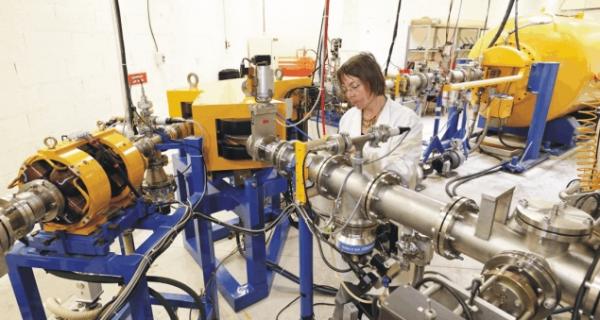
La microsonde nucléaire est un outil d’analyse non destructif permettant la caractérisation élémentaire d’échantillons de nature solide de provenances très diverses, et ce à l’échelle du micromètre : interfaces et grains de matériaux de synthèse, monocouches cellulaires, inclusions dans des échantillons géologiques terrestres et extra-terrestres…
L'accès à l'appareil se fait par "demande d'expérience soumise à expertise et évaluation par un comité".
Les rayons X, rayonnement électromagnétique au delà de l'ultra-violet lointain, couvrent une gamme de longueur d'onde autour du dixième de nanomètre. Cette distance est de l'ordre de la distance entre atomes dans la matière condensée. Ainsi les rayons X peuvent interagir avec ces atomes (diffraction) ou les électrons (diffusion).

x-rays, electromagnetic radiation beyond the remote ultraviolet ray, cover a range wavelength around the tenth of nanometer. This distance is about the distance between atoms in the condensed matter. The diffraction of x-rays thus makes it possible to probe the matter, and to obtain information on the structure, the order and the composition of materials.

The nuclear microprobe is a non-destructive analysis tool allowing elemental characterization at the micrometer scale of solid samples of diverse origins: interfaces and grains of synthesized materials, cellular monolayers, inclusions in terrestrial and extraterrestrial geologic samples…
The method relies on the detection and spectrometry of emitted radiations by the interaction of a light ion micro-beam with the atoms composing the major elements or traces of the samples.
Dès le début de la découverte de la radioactivité, les chercheurs se sont intéressés aux effets chimiques provoqués par les rayonnements. Malgré la faiblesse des sources radioactives de l'époque qui rendait les expériences quantitatives pratiquement impossibles, on a trouvé que l'eau irradiée par les rayons a du radium se décomposait en formant de l'hydrogène et de l'oxygène.

The LAPA developed for re-corroding archaeological systems, originals setups, allowing to continue century old corrosion process in the lab and under labeled environments.
One way to get reliable knowledge about long term corrosion is to perform re-corrosion experiments. From natural sites, known for their chemical stability for years, corroded samples are picked up and then submitted to experiments.
The Raman microspectrometry is an analytical device of primary importance to characterize the crystalline structure of materials. It is a complementary technique to micro X-ray diffraction. This technique is well suited to characterize and determine the distribution of the phases formed in the corrosion layers of iron or steels during very long periods in various environments. Iron oxides, oxy-hydroxides and carbonates are the main phases encountered in these systems.