
A comparison of lead and cadmium accumulation in renal epithelial cultured cells after acute intoxication
M. Carrière, L. Avoscan, H. Khodja, F. Carrot, B. Gouget

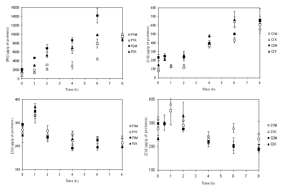
Figure 1. Uptake of Cd or Pb by MDCK and NRK-52E cells vs. intracellular Zn content as a function of time. The cells were exposed to 20 (C1M for the MDCK cells, C1K for the NRK-52E cells) and 50 (C2M and C2K) µM of Cd and 80 (P1M for the MDCK cells, P1K for the NRK-52E cells) and 200 (P2M and P2K) µM of Pb. Each point represents the mean SD of three replicate samples.
Pb or Cd overexposure results in acute nephrotoxicity mostly consisting in proximal renal tubular dysfunction and accumulation. Acute Pb nephropathy is limited to functional and morphological changes in cells which are reversible by treatment with a chelating agent. Nevertheless, progression from acute reversible to chronic irreversible Pb nephropathy has not been clearly explained. Cd nephrotoxicity is probably not reversible and results from a gradual accumulation in the renal cortex. There is no effective agent for chelating cadmium from the kidney. After Pb or Cd acute intoxications, a metal-protein complex is formed in renal cells that sequesters the metal in a non-toxic form. The Pb-protein complex is insoluble and can be observed microscopically, but little is known about the nature of the protein. On the contrary, the soluble Cd-protein complex, cadmium-metallothionein is quite well documented. However, even if several hypotheses have been proposed, the mechanism of its protective effect remains unclear. The use of cell culture systems to study the cellular uptake, metabolism and toxicity of chemicals has improved the understanding of mechanisms of metals cellular effects. Kidney epithelial cell lines can be used for direct comparison of cytotoxicity of metals. In this study, Cd and Pb toxicity to renal cells was investigated on cell lines representative of the distal or proximal tubules of the nephron. After cells exposure to different concentrations of metal for various times, morphological changes on treated cells were observed and intracellular concentrations of toxic metals versus biological elements were measured by inductively coupled plasma-mass spectrometer analysis. Results on Cd and Pb toxicity to these renal epithelial cells will be presented. Further study on metal speciation should permit to identify metal-protein complexes of Pb or Cd in cells and to better understand their effects against metal stress.
#426 - Màj : 25/03/2005
• ![]() Physics and chemistry for life sciences and the environment › Human and Environmental Toxicology
Physics and chemistry for life sciences and the environment › Human and Environmental Toxicology
• Laboratoire Pierre Süe UMR 9956 CEA-CNRS • Pierre Süe Laboratory UMR 9956 CEA-CNRS
