Laboratoire Léon Brillouin
UMR12 CEA-CNRS, Bât. 563 CEA Saclay
91191 Gif sur Yvette Cedex, France
+33-169085241 llb-sec@cea.fr

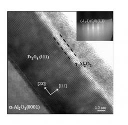
Figure 1: HRTEM micrograph of the Fe3O4 (15 nm)/γ-Al2O3 (1.5 nm) bilayer. The similarity of the two lattices (spinel) demonstrates that the Al2O3 layer grows in the γ phase (Coll. P. Warin, P. Bayle-Guillemaud DRFMC-SP2M). Inset displays the RHEED pattern along the [1-100] direction of the crystalline γ-Al2O3 layer at the end of the growth.
At first we have studied the growth of fully epitaxial Fe3O4(111)/γ-Al2O3(111) bilayers to be included in a MTJ. Epitaxial growth by molecular beam epitaxy (MBE) of a γ-Al2O3 ultra-thin layer (a few nanometers) on top of a magnetite electrode was achieved by evaporation of metallic aluminium from a Knudsen effusion cell under atomic oxygen. The RHEED diffraction patterns (inset of figure 1) recording during the growth demonstrated the two-dimensional growth mode of Al2O3. In situ Auger electron spectroscopy (AES) and X-ray photoelectron spectroscopy (XPS) measurements (Fe 2p and Al 2p core levels) have also been performed in order to check the oxidation state of Fe at the Fe3O4/Al2O3 interface and the full homogeneous oxidation of aluminium. In appropriate growth conditions (Al flux, atomic oxygen pressure), we were able to obtain a stoichiometric Fe3O4/Al2O3 interface [1,2]. The magnetic properties of these stoichiometric bilayers, obtained by VSM magnetometry and XMCD analyses (not shown here), exhibited the same magnetic behavior (remanence, coercivity, Verwey transition, magnetic moment) as the corresponding uncovered Fe3O4 layers. The HRTEM cross section in figure 1 confirmed the very good epitaxy of the Fe3O4(111)/γ-Al2O3(111) system. Finally, the insulating properties of the γ-Al2O3(111) ultra-thin layers have been measured using a conductive tip AFM (Coll. UMR CNRS/Thales) validating the tunnel barriers properties [1,2]. No pinholes have been detected, demonstrating that the Fe3O4 layer is entirely covered by a 1.5 nm thick Al2O3 layer.
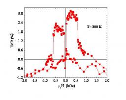
Figure 3: Room temperature magnetoresistance (TMR) of a 96 μm2 Fe3O4 (25 nm)/γ-Al2O3 (2 nm)/Co (15 nm) magnetic tunnel junction (Coll. R. Mattana, P. Seneor, K. Bouzehouane, F. Petroff, UMR CNRS/Thalès).
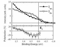
Finally, MTJs were prepared by covering the Fe3O4/γ-Al2O3 bilayers with an iron or cobalt counter-electrode. The magnetic hysteresis loop of a Fe3O4 (25 nm)/γ-Al2O3(3 nm)/Co(15 nm) trilayer shows a clear magnetic decoupling between the two ferromagnetic electrodes. Figure 3 displays a TMR curve obtained on a 96 mm2 MTJ (coll. UMR CNRS/Thales) at room temperature. The first abrupt switching around 50 Oe corresponds to the cobalt layer and the second switching around 600 Oe is related to Fe3O4 with probably a slight magnetic coupling. The first TMR measured, normalized with respect to R(μ0H= 0), is around +3 % at room temperature [1, 5] and does not depend on temperature in the range 130 K–300 K. Considering the Jullière’s model, this positive TMR seems to be in contradiction with the negative spin polarization of the Fe3O4/γ-Al2O3 determined by spin polarized photoemission and the positive spin polarization of the Co/Al2O3 interface generally observed. Different studies are in progress in order to understand these first results (effects of APBs, lithography, …) and to optimize the absolute value of the TMR.
•  Future optics and electronics › Nanomagnetism, spintronic and multiferroic materials
• Service de Physique et Chimie des Surfaces et des Interfaces • Laboratory of Physics and Chemistry of Surfaces and Interfaces
• Laboratoire des Interfaces et Surfaces d'oxydes (LISO) • Laboratory of Oxide Surfaces and Interfaces
