
Service de Physique de l'Etat Condensé


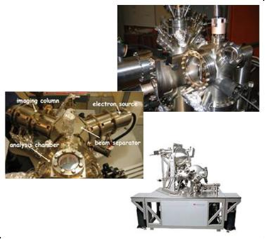
Microscope STM, LEEM-PEEM et MesoXcope de la plateforme de caractérisation UHV de l'IRAMIS.
Identifier proprement la structure, la caractérisation chimique et les propriétés physiques des objets à l'échelle atomique et moléculaire demande le plus souvent de travailler sous ultra-vide, afin d'éviter toute contamination et sans interférence avec un dispositif de protection (surcouche, encapsulation, …).
L'IRAMIS/SPEC implanté sur le Centre de Saclay dispose d'un ensemble d'appareils de caractérisation :
Cet ensemble est accessible pour la réalisation de projets scientifiques et aux entreprises, par l'intermédiaire de collaborations financées avec les équipes spécialistes de chacun de ces instruments.
Description technique et pages scientifiques associées.
Contact : Luc Barbier.
Properly identify the structure, the chemistry and the physical properties of objects at the atomic and molecular scale requires studies under ultra high vacuum to avoid contamination and any interference with a protective device (overlay, encapsulation , ...).
IRAMIS / SPEC located on the CEA Saclay Center offers a set of such characterization equipments:
This set is available for the realization of technical and scientific projects, through funded collaborations with the specialist teams in charge of each of these instruments.
- Technical description and associated scientific pages.
Contact : Luc Barbier.
• ![]() Physique, chimie, nanosciences et matériaux autour des grands instruments
Physique, chimie, nanosciences et matériaux autour des grands instruments
• ![]() Institut Rayonnement Matière de Saclay • Laboratory of Physics and Chemistry of Surfaces and Interfaces • UMR 3680 - Service de Physique de l'Etat Condensé (SPEC) • Service de Physique et Chimie des Surfaces et des Interfaces
Institut Rayonnement Matière de Saclay • Laboratory of Physics and Chemistry of Surfaces and Interfaces • UMR 3680 - Service de Physique de l'Etat Condensé (SPEC) • Service de Physique et Chimie des Surfaces et des Interfaces
• Microscopies à sonde locale • Microscopies électroniques TEM, MEB et LEEM/PEEM
