


Invité par Nick BARRETT,
Abstract :
X-ray photoelectron spectroscopy (XPS) is widely utilized for the characterization of surfaces and interfaces, owing to its chemical and surface sensitivity. Ferroelectrics are intensely studied materials nowadays, but only recently models were proposed for charge compensation of the depolarization field [1]. Recently it was shown that high resolution XPS is able to quantify band bending at surfaces and interfaces [2-6]. Core level shifts are sensitive to the conduction and valence band bending at interfaces, which is of particular interest for ferroelectrics (Fig. 1) and for metal/ferroelectric interfaces. In this work we will review recent results obtained by using XPS to quantify band bendings at metal-semiconductor interfaces [3], at free ferroelectric surfaces [2,4], and at metal-ferroelectric interfaces [5], evidencing new phenomena such as competing Schottky barrier and over-compensation by electrons provided by the metal (Fig. 2) [6], or preferential adsorption of polar molecules onto areas with well-defined out-of-plane polarization. New functionalities of ferroelectrics start to be exploited in surface chemistry and catalysis. Fig. 3 demonstrates the interplay between surface core level shifts and the molecular coverage for CO adsorbed on Pb(Zr,Ti)O3.
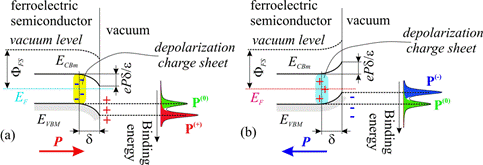
Figure 1: Band bendings at free ferroelectric surfaces and their effects on the core level shifts: (a) outwards polarization; (b) inwards polarization.
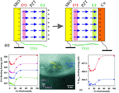
Figure 2: Depolarization mechanism proposed to explain the vanishing of the surface band bendings when Cu layers are grounded, together with observed core level shifts and evidence of Cu nanoparticle formation by high resolution transmission electron microscopy.
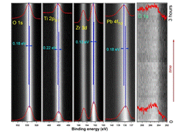
Figure 3: High resolution XPS spectra obtained at SuperESCA on an atomically clean PZT(001) single crystal layer where intially 6000 L of CO were dosed. Synchrotron radiation promotes CO desorption, and core levels are shifted towards higher binding energies.
[1] L. Pintilie and M. Alexe, J. Appl. Phys. 98, 124103 (2005).
[2] F. Chen and A. Klein, Phys. Rev. B 86, 094105 (2012).
[3] N.G. Apostol, L.E. Stoflea, G.A. Lungu, C. Chirila, L. Trupina, R.F. Negrea, C. Ghica, L. Pintilie, C.M. Teodorescu, Appl. Surf. Sci. 273, 415-425 (2013).
[4] N.G. Apostol, L.E. Stoflea, G.A. Lungu, C.A. Tache, L. Pintilie, C.M. Teodorescu, Mater. Sci. Eng. B 178, 1317-1322 (2013).
[5] N.G. Apostol, L.E. Stoflea, G.A. Lungu, L.C. Tanase, C. Chirila, L. Frunza, L. Pintilie, C.M. Teodorescu, Thin Solid Films 545, 13-21 (2013).
[6] N.G. Apostol, L.E. Stoflea, C. Chirila, L. Trupina, R. Negrea, L. Pintilie, C.M. Teodorescu, J. Mater. Sci., in press (2014).
http://old.infim.ro/~lab150/highlights/html/dr__cr...
