


Pages scientifiques 2015

A plasma mirror is a dense plasma created at the surface of an initially solid target when this target is irradiated by an intense femtosecond laser pulse. Due to its high density (≈10 23 electrons/cm3), it efficiently reflects the incident laser field. In addition, given the brevity of the laser pulse, its interface remains optically-flat during the interaction: plasma mirrors thus specularly reflect the incident laser beam, and can be viewed as high-quality mirrors suitable for UHI beams [Kap91Dou04,Tha07] (Fig.1), with many potential applications (Applications of plasma mirrors). They are also ideal systems to investigate the fundamental physics of UHI interactions (Fundamental interest of plasma mirrors). The Physics at High Intensity group has made major contributions to the investigation of plasma mirrors in the last decade (Results of the PHI on this topic).

Fig. 1: Physics of plasma mirrors. When exposed to UHI laser fields, plasma mirrors specularly reflect the laser with a reflectivity higher than 70% (bottom left image). Their highly-nonlinear coupling with the field (upper left image, from PIC simulations) also leads to the emission of beams of high-order harmonics and relativistic electrons. Typical spatial patterns of these beams are shown in the central images, and their typical energy spectra in the rightmost images (from experiments with the UHI100 laser at I=3.1019 W/cm2).
[Dou04] Doumy et al, Phys. Rev. E 69, 026402 (2004)
[Kap91] Kapteyn et al, Opt. Lett. 16, 490 (1991)
[Tha07] Thaury et al, Nature Physics 3, 424 - 429 (2007)
Magnetophysiology is the study of the magnetic fields generated by living organisms. In the case of ionic currents flowing into neurons or muscles, the generated magnetic signal is in the picotesla (10-12T) to nanotesla (10-9T) range, i.e. thousands to millions times smaller than the Earth field. SQUIDS are commonly used to sense these very weak magnetic fields [1], [2]; however, as they have to be kept at liquid helium temperature, they use at cellular level measurements can be complicated and impossible for microscale in vivo applications.
In this project, the probes developed are called “Magnetrodes” as a magnetic equivalent of electrodes. They are based on spin electronics which offer the possibility to be miniaturized and sensitive enough (at room temperature) to detect the very weak magnetic signature of neural activity at cellular level .
At CEA-Saclay, we develop two kinds of probes to record the magnetic signature at mesoscale and microscale for in vitro and in vivo experimentations.
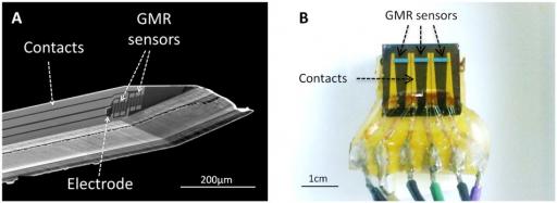
At microscale, sharp probes have been designed in a needle shape comparable to electrode design used for electrophysiology. These probes are used as deep sensors to perform localized magnetic recordings of the local neuronal activity. Sharps probes contain one or two GMR elements close to the tip of the probe, enabling gradiometric measurements. An electrode can be located next to the GMR sensors to have a local reference of the LFPs. They are fabricated on very thin Silicon substrate (from 100 to 200 µm) in order to be as less invasive as possible.
At mesoscale, planar probes are used to measure the magnetic field created at the surface of the muscular fibers or at the surface of the cerebral cortex. Planar probes contain a rather large GMR element deposited on a conventional 700µm-thick silicon substrate.
Our measurements are based on three different setups:
- Experimentations in vitro with the muscle fibers of the mouse soleus, where the goal is to test the magnetrode technology with a relatively simple model of muscular action potential propagation.
- Experimentations in vitro with slices of brain hippocampus, where the neurons are parallel arranged and all the somata are lying on a same layer providing a summation of the magnetic field.
- Experimentation in vivo on the visual cortex, where evoked neuronal responses are induced by direct visual stimulus.
The development of highly controllable oxide thin films growth and lithography techniques makes it possible to realize genuine materials stacking and nanostructures dedicated to spintronics applications like magnetic tunnel junctions, spin filters and more recently multiferroics. A renewed interest on ferroelectric materials has emerged for the development of nonvolatile random access memories and multiferroic materials. In multiferroics several ferroic orders can coexist and be eventually coupled, as for example ferroelectric, ferroelastic and/or ferro- (or antiferro-) magnetic long-range orders. The multiferroics class of materials has commonly been extended to anti-ferroics which show antiferromagnetic and antiferroelectric behaviors. Unfortunately, single phase multiferroics with ordering temperatures above 300 K are very seldom and artificial multiferroics are actively sought after. Defining and tailoring new operational multiferroic materials has, in recent years, emerged as an important topic in modern spintronics driven by numerous potential applications and fundamental physics issues such as the coupling between ferroelectric and magnetic orders and domain structures. This opens exciting new perspectives like the control of the magnetic state of an individual device by an electric field (and/or the control of the ferroelectric state by a magnetic field).
Artificial multiferroics can be realized following several routes: (i) doping a ferroelectric material with ferromagnetic ions, (ii) co-deposition and thermal treatments taking advantage of phase separation thermodynamics, (iii) the combination of ferroelectric and ferromagnetic materials in multilayered or nanostructured systems. The coupling between ferroelectricity and magnetism through interface bonding has been predicted theoretically for ferromagnetic/ferroelectric interfaces and was indeed observed in some systems. Additional magnetic exchange coupling using antiferromagnetic layer and strain coupling can be exploited as well.
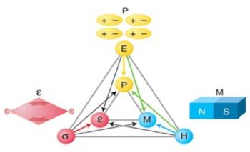
The functioning principle of multiferroics dedicated to spintronics relies on a subtle interplay between structural strain, magnetization and electric polarization (figure 1). A fundamental understanding of multiferroic materials necessarily needs to address all these properties at the same time which requires the elaboration of single crystalline multiferroic model samples and experimental techniques or tools able to address each every aspect.
The laboratory has acquired expertise in growing epitaxial oxide thin films by atomic oxygen plasma assisted molecular beam epitaxy (AO-MBE). In particular of the ferroelectric perovskite barium titanate (BaTiO3) and ferrimagnetic spinel ferrites (ex. CoFe2O4) thin films, the former is an excellent and well-known ferroelectric material that displays remnant electric polarization higher than bulk crystals and the later class of materials presents a wide range of magnetic properties at room temperature.
In addition to classical laboratory characterizations, the model multiferroics materials are studied within a wide range of pertinent additional synchrotron radiation techniques allowing to access the structure (X-ray diffraction and absorption), the element specific or average magnetic properties (magnetic dichroism, spectromicroscopy) and electrical and magnetic domain structure (spectromicroscopy) that allows determining the electronic and crystalline structures as well as the ferroic orders.
12-18 months Postdoctoral Position in CEA-Saclay (France). Starting Falls 2015
Contact: sebastien.aumaitre@cea.fr
 |
 |
Energy storage optimization in power grid with intermittent renewable power sources

