


Epitaxial growth of nanometric Fe3O4 thin films

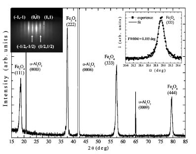
X-ray diffraction pattern of a 15 nm thick Fe3O4(111) film. Right inset: rocking curve of the (333) peak. Left inset: RHEED pattern of the Fe3O4 thin film.
Magnetite Fe3O4 is a very attractive oxide for applications in spin electronic devices because this material has a high Curie temperature (Tc= 860 K)and is expected to be an half-metal (i.e. a material which is a conductor for one spin direction and an insulator for the other one).
In this framework, a team of the SPCSI has developped a molecular beam epitaxy (MBE) setup assited by a radio frequency plasma source specially dedicated to the growth of iron oxides layers on alumina substrates. The thin films are characterized during and after the growth by different techniques: low energy (LEED) and high energy (RHEED) electron diffraction, X-ray photoemission (XPS) and Auger electron (AES) spectroscopy.
We are able to realize the epitaxial growth of magnetite thin films with high structural properties as shown in this transmission electron microscopy (TEM) picture. However we can also observe the presence of structural defects such as antiphase boundaries (APBs).
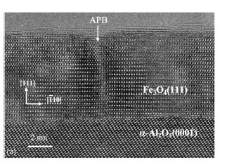
Cross-sectional HRTEM image of a 15 nm thick Fe3O4(111) thin film with the presence of an APB grown on an alumina substrate (Coll. P. Bayle-Guillemaud,DSM/DRFMC/SP2M).
#478 - Last update : 07/18 2005
• ![]() Future optics and electronics › Nanomagnetism, spintronic and multiferroic materials
Future optics and electronics › Nanomagnetism, spintronic and multiferroic materials
• Laboratory of Physics and Chemistry of Surfaces and Interfaces
