


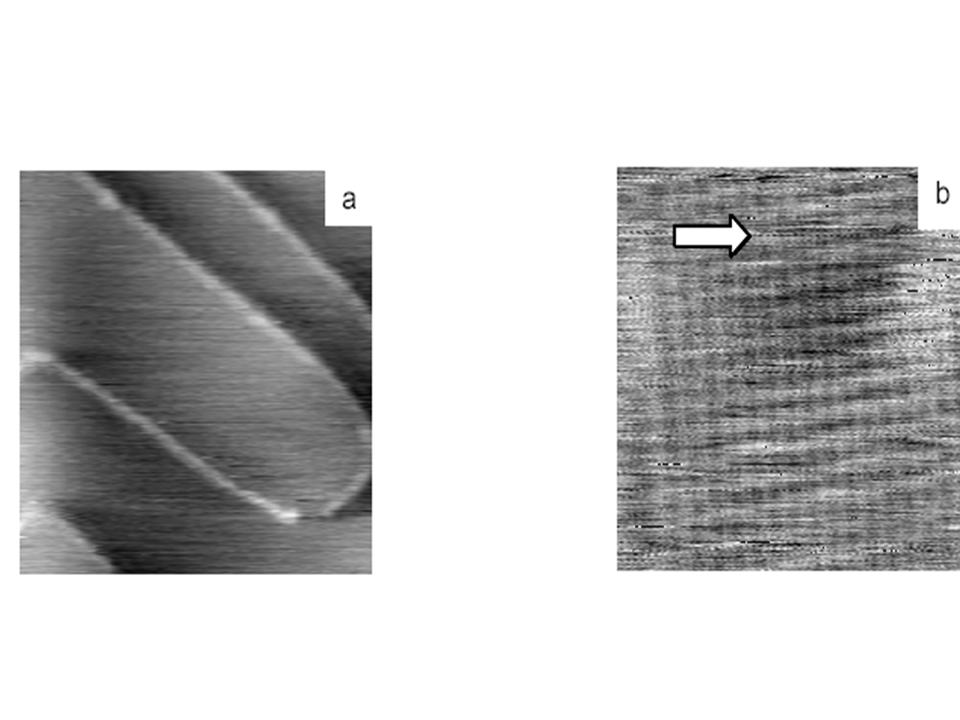
Figure 1 : NC AFM micrographs of a (001) KBr cleaved surface obtained in UHV. In fig1 a, large terraces and monoatomic steps are imaged. (Size 120x120nm²) The square atom packing (atomic corrugation 50pm) of the surface is shown in fig b. (size 5 x 5nm²)
Achieving NC-AFM atomic resolution images on an insulator depends mainly on the conjunction of three constituents: a sharp tip, a suitable sample - i.e atomically flat and to a certain extent free of electric charges -, and a low noise, stable microscope. While requiring some patience, testing the first two conditions is experimentally accessible.
In order to test the microscope, we measured the noise of the optical detection and that of our custom demodulator as a function of the cantilever amplitude. For the noise at the output of the phase detector, two different sources were used: first, the experimental optical deviation signal induced by the harmonic motion of the cantilever, and second, a dummy sinusoidal signal produced by a reference generator noise. We tried to filter the signal but we finally ended up replacing the light source.
We could find an IR Laser diode intended for aerospace applications and we have developed a dedicated optical system. After an adjustment period, the new source could clearly outperform the former one while allowing to keep the same structure for the microscope stage. Also gains in term of thermal stability could be achieved as demonstrated by the drift in the cantilever eigenfrequency, which dropped from 70Hz to 2-3Hz.
Several crystal surfaces were examined. On a flamed surface of Au(111) the presence of a herringbone structure is clearly visible and presents corrugation of a few tens of picometers. We have also tested atomic resolution on a KBr(001) sample, cleaved under a nitrogen atmosphere. The sample was baked in-situ (one hour) to remove the contamination layer and the residual charges as proposed by Hoffmann.[1] Fig.1 presents two AFM micrographs obtained at different scales. While flat terraces and steps are clearly seen in Fig. 1a, a square lattice is observed in Fig. 1b. In this image, no point defect is visible; however, a true atomic resolution can be inferred from the fact that the presence of a bump on the upper left of the image (arrow) slightly distorts the atomic lattice.
• Service de Physique et Chimie des Surfaces et des Interfaces
