Les sources d’électrons pulsées représentent une alternative intéressante aux sources de photons X pulsées basées sur des systèmes laser de haute intensité.
Nous décrirons les méthodes actuelles de génération de paquets d’électrons ainsi que les techniques de caractérisation des paquets. Les résolutions temporelles et spatiales ultimes qu’il est possible d’atteindre à ce jour seront discutées. Nous exposerons les résultats obtenus lors de notre dernière campagne de mesure sur le serveur ELYSE de l’Université Paris-Sud en 2014, en collaboration avec une équipe de l’ISMO et la start-up ITEOX. Finalement, quelques applications seront mises en perspective.
Coherent diffractive imaging is a recent imaging technique that offers spatial resolutions of the order of the wavelength, thanks to the fact that it does not require any potentially aberrating optical elements. We recently demonstrated sub 100-nm resolution using XUV radiation created by infrared laser high order harmonic generation (HHG). However, the resolution is usually limited by the source spectral width. We propose a new lensless holographic imaging scheme that gives the ability to exploit the spectral and temporal properties of HHG emission. Through a clever positioning of the holographic reference, we demonstrate the possibility either to combine the spatial and spectral resolutions in a single laser pulse, or to achieve nanometer spatial or sub-femtosecond temporal resolutions.

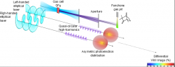
Due to their considerable energy, extreme ultraviolet photons ionize all molecules, regardless of the details of their energy structure. For this reason ultrashort light pulses in this spectral range are second to none to probe photochemical processes. In particular they give access to information on the reaction intermediates, which are ephemeral structures whose spectroscopic properties are usually out of reach. Through a collaboration between the Laboratory interactions, dynamic and lasers - LIDyL (CEA), the intense laser center for applications - CELIA (CNRS / CEA / Univ Bordeaux.), The SOLEIL synchrotron, and the Laboratory collisions, aggregates, reactivity - LCAR (CNRS / Univ. Toulouse 3) we have developed a new laboratory scale source that delivers bright coherent ultrashort and quasi-circularly polarized pulses in the extreme ultraviolet. For this, we used resonant high order harmonic generation in a gas submitted to an intense laser pulse. Today, such a circularly polarized light is produced in this range of radiation by just a few large scale facilities such as synchrotrons and, with the notable exception of a few free electron lasers, only quasi-continuously. Specific polarization properties of this new source presage pump probe studies of processes occurring in chiral molecules, that is to say molecules that are not their own mirror image. The ubiquitous role of these molecules in organic chemistry and biology suggests many applications.
http://www.nature.com/nphoton/journal/v9/n2/full/nphoton.2014.314.html
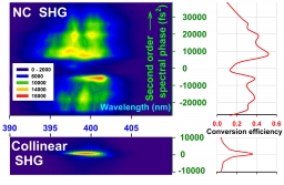
We report on a 400 nm broad-band type I frequency doubling in a non-collinear geometry with pulse front tilted and chirped femtosecond pulses (l0 = 800 nm, FTL pulse duration ~ 45 fs). With moderate power densities (2 to 10 GW/cm2) thus avoiding higher-order nonlinear phenomena, the energy conversion efficiency was up to 65%. Second-harmonic pulses of Fourier transform limited pulse duration shorter than the fundamental wave were generated, exhibiting a good beam quality and no pulse-front tilt. High energy (20 mJ/pulse) was produced in a 40 mm diameter and 6 mm thick LBO crystal. To the best of our knowledge, this is the first demonstration of this optical configuration with sub 100 fs pulses. A good agreement between experimental results and simulations is obtained.
|
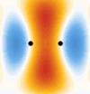
Visualizing the motion of electrons in matter requires both a spatial resolution at the Angstrom scale and a temporal resolution at the attosecond scale (1 as = 10-18 s). Such an "ultra-fast camera" opening a path towards "viewing the electrons" is demonstrated: it allows imaging molecular orbitals using the ultra-short attosecond emission from this orbital in an intense laser field. In a close collaboration, scientists from IRAMIS-SPAM, LCPMR of the Univ. Paris 6 and from CNRS (UMR 7614) have shown, in experiments realized within the CEA-Saclay, the possibility of an "Attosecond-Angstrom" imaging in the case of the nitrogen molecule N2. |
In photography, the scattered light from an illuminated object is recorded with a detector and one get an image of it. If the image is formed with an objective, the optics imposes many limitations (resolution, aberrations ...). To achieve the ultimate resolution, spatially (function of the wavelength of the radiation used) and temporally (function of the "flash" duration), one possible technique (without any optics) is the coherent diffraction. Using a coherent beam like a laser to illuminate the object, a signal modulation due to interference is present and allows digitally reconstructing the exact image of the object with an unprecedented precision. To achieve nanometer or even atomic resolution, we therefore enlighten with a beam of coherent X-rays (radiation laser wavelength nanometer) and record the image. The usually low average illumination requires long accumulations over several laser shots. Recent advances have yielded images with a single shot femtosecond (10-15 s) from a laser laboratory, opening the way for time resolved studies.
For regular arrangements of elementary objects, the Bragg diffraction of X-rays is a powerful technique for characterization of matter at the atomic scale. It is the primary tool for crystallography. The information contained in the Bragg diffraction is rich: if a is the characteristic size of the elementary object, the Bragg peaks are spaced by 1/a in reciprocal space. However, some information is lost: indeed, the maximum frequency at which the diffraction pattern can be sampled is less than the Nyquist frequency (2a). In particular, if the elementary object has an amplitude and a phase, the phase is lost in the Bragg diffraction.

G. Lambert1,2,3, T. Hara2,4, D. Garzella1, T. Tanikawa2, M. Labat1,3, B. Carre1, H. Kitamura2,4, T. Shintake2,4, M. Bougeard1, S. Inoue4, Y. Tanaka2,4, P. Salieres1, H. Merdji1, O. Chubar3, O. Gobert1, K. Tahara2, M.-E. Couprie3
1Service des Photons, Atomes et Molécules, DSM/DRECAM, CEA-Saclay, 91191 Gif-sur-Yvette, France
2RIKEN SPring-8 Centre, Harima Institute, 1-1-1, Kouto, Sayo-cho, Sayo-gun, Hyogo 679-5148, Japan
3Groupe Magnétisme et Insertion, Synchrotron Soleil, L'Orme des Merisiers, Saint Aubin, 91192 Gif-sur-Yvette, France
4XFEL Project Head Office/RIKEN, 1-1-1, Kouto, Sayo-cho, Sayo-gun, Hyogo 679-5148, Japan
The SNOM (Scanning Near-field Optical Microscopy) allows to reach a spatial resolution well below the wavelength of the light used. Indeed, thanks to the "near-field" illumination (when the distance between the object and the source is much less than the wavelength) it becomes possible to avoid the diffraction inherent to every optical system. Measurement of the evanescent field at the back side of a total-reflection interface allows obtaining a SNOM image with a resolution well below one micrometer.